
Group Leader Joaquín Arribas Postdoctoral Fellows Marta Lalinde, Vanesa Nogales, Macarena Román Graduate Students Ariadna Grinyó, Jade Liu Technicians Marta Escorihuela, Susana Maqueda, Sandra Perez Visiting Scientists Marta Bort, Santiago Duro, Judit Gago, Pau García,
Pau Garcíal
Growth Factors Group
Our group continues advancing along two main research lines: the development of novel therapies targeting the tyrosine kinase receptor HER2, and the characterization of therapy-induced cellular senescence during cancer progression and treatment.
In 2024, we completed the development of a next-generation chimeric antigen receptor (CAR) T cell therapy with remarkable anti-tumor activity. This novel strategy involves T cells transduced with a retroviral vector encoding a second-generation CAR targeting the tumor-specific antigen p95HER2, along with a bispecific T cell engager that binds selectively to HER2-overexpressing cells. These engineered T cells, which we have named p95HER2.CAR-TECH2Me, demonstrated strong efficacy, achieving complete responses in breast, gastric, and pancreatic cancer PDX models. Importantly, they appear to be safe, as they do not affect cells expressing normal levels of HER2. These findings pave the way for the clinical testing of p95HER2.CAR-TECH2Me T cells (Román et al. Nat Commun. 2024).
In collaboration with Manuel Serrano’s group, we explored the role of cellular senescence in cancer. We found that PD-L2 (programmed cell death ligand 2), a ligand of the PD receptor, is frequently expressed by senescent cells. Various chemotherapy regimens induced cellular senescence and, consequently, an immunosuppressive tumor microenvironment in breast cancers. Treatment with anti-PD-L2 antibodies resulted in the rapid clearance of senescent cells and subsequent infiltration of anti-tumorigenic T cells. This led to durable responses, supporting the conclusion that combining chemotherapy with anti-PD-L2 is significantly more effective than chemotherapy alone (Chaib et al. Nat Cancer. 2024).
We have expanded our platform of HER2-positive patient-derived xenograft (PDX) models to include tumors from different origins, including endometrial, hepatobiliary, and pancreatic cancers (Yáñez-Bartolomé et al. Methods Mol Biol, 2024). Our ultimate goal is to apply our novel anti-HER2 therapies across various tumor types. This expanded PDX platform will be instrumental for advancing our research on HER2+ cancers and fostering collaboration with other VHIO groups.
Our PDX platform has also enabled multiple intra- and extramural collaborations. We demonstrated that PARP inhibition elicits a potent and durable antitumor response in BRCA1/2-associated patient-derived cancer models (Herencia-Ropero et al. Genome Med. 2024). In addition, we showed that inhibitors of ERK1/2 dimerization impair cancer cell motility (Fuente-Vivas et al. Mol Oncol. 2024).
- Generate and characterize next generation chimeric antigen receptor (CAR)-T cells against HER2-positive tumors.
- Develop novel strategies to deplete deleterious senescent cells.
- Characterize new mechanisms of resistance to targeted therapies.
- The production of p95HER2.CAR-TECH2Me T cells for a phase I clinical trial.
- Blocking PD-L2 is a novel anti-tumor strategy to complement different chemotherapeutic regimes.
- Test p95HER2.CAR-TECH2Me T cells in a phase I clinical trial.
- The development of novel senolytic strategies
- Generation of immunocompetent mouse models to evlaute anti-HER2 therapies.
- Román Alonso M, Grinyó-Escuer A, Duro-Sánchez S, Rius-Ruiz I, Bort-Brusca M, Escorihuela M, Maqueda-Marcos S, Pérez-Ramos S, Gago J, Nogales V, Espinosa-Bravo M, Peg V, Escrivá-de-Romaní S, Foradada L, Soucek L, Braña I, Galvao V, Martín-Lluesma S, Moessner E, Klein C, Garralda E, Saura C, Arribas J. Generation of chimeric antigen receptor T cells targeting p95HER2 in solid tumors. Nat Commun. 2024 Nov 18;15(1):9589.
- Title: SGR 2017-2019 Funding agency: Agència de Gestió d’Ajuts Universitaris i de Recerca AGAUR/Fons Europeus de Desenvolupament Regional (FEDER) 2014-2020 Reference: 2017 SGR 532 Duration: Jan 2018 – Dec 2021
- Title: Generation of CAR T cells against FGFR2 S252W in Endometrial Cancer Funding agency: Fundación La Caixa Duration: 1 Jan 2025 – 31 Dec 2025
- Title: Redirection of T cells against HER2-driven tumors Funding agency: Asociación Española Contra el Cáncer (AECC) Reference: GCAEC19017ARRI Duration: Nov 2019 - Nov 2024
- Title: Immune Senolysis Against Breast Cancer Funding agency: Breast Cancer Research Foundation (BCRF) Reference: BCRF-24-008 Duration: October 2024 – October 2025.
- Title: Overcoming Drug Resistance in HER2-Positive Breast Cancer Funding agency: National Institutes of Health Reference: R01CA244601-01 Duration: Jan 2020 – Jan 2025
- Title: EDIReX: EurOPDX Distributed Infrastructure for Research on patient-derived cancer Xenografts Funding agency: European Union. Horizon 2020 call H2020-INFRAIA-2016-2017 “Integrating activities for starting communities" Reference: EDIReX Duration: Oct 2017-Oct 2022
- Román Alonso M, Grinyó-Escuer A, Duro-Sánchez S, Rius-Ruiz I, Bort-Brusca M, Escorihuela M, Maqueda-Marcos S, Pérez-Ramos S, Gago J, Nogales V, Espinosa-Bravo M, Peg V, Escrivá-de-Romaní S, Foradada L, Soucek L, Braña I, Galvao V, Martín-Lluesma S, Moessner E, Klein C, Garralda E, Saura C, Arribas J. Generation of chimeric antigen receptor T cells targeting p95HER2 in solid tumors. Nat Commun. 2024 Nov 18;15(1):9589. doi: 10.1038/s41467-024-53265-7.
- Chaib S, López-Domínguez JA, Lalinde-Gutiérrez M, Prats N, Marin I, Boix O, García-Garijo A, Meyer K, Muñoz MI, Aguilera M, Mateo L, Stephan-Otto Attolini C, Llanos S, Pérez-Ramos S, Escorihuela M, Al-Shahrour F, Cash TP, Tchkonia T, Kirkland JL, Abad M, Gros A, Arribas J, Serrano M. The efficacy of chemotherapy is limited by intratumoral senescent cells expressing PD-L2. Nat Cancer. 2024 Mar;5(3):448-462. doi: 10.1038/s43018-023-00712-x. Epub 2024 Jan 24.
- Herencia-Ropero A, Llop-Guevara A, Staniszewska AD, Domènech-Vivó J, García-Galea E, Moles-Fernández A, Pedretti F, Domènech H, Rodríguez O, Guzmán M, Arenas EJ, Verdaguer H, Calero-Nieto FJ, Talbot S, Tobalina L, Leo E, Lau A, Nuciforo P, Dienstmann R, Macarulla T, Arribas J, Díez O, Gutiérrez-Enríquez S, Forment JV, O'Connor MJ, Albertella M, Balmaña J, Serra V. The PARP1 selective inhibitor saruparib (AZD5305) elicits potent and durable antitumor activity in patient-derived BRCA1/2-associated cancer models. Genome Med. 2024 Aug 26;16(1):107. doi: 10.1186/s13073-024-01370-z.
- Serra-Bardenys G, Blanco E, Escudero-Iriarte C, Serra-Camprubí Q, Querol J, Pascual-Reguant L, Morancho B, Escorihuela M, Tissera NS, Sabé A, Martín L, Segura-Bayona S, Verde G, Aiese Cigliano R, Millanes-Romero A, Jerónimo C, Cebrià-Costa JP, Nuciforo P, Simonetti S, Viaplana C, Dienstmann R, Oliveira M, Peg V, Stracker TH, Arribas J, Canals F, Villanueva J, Di Croce L, García de Herreros A, Tian TV, Peiró S. LOXL2-mediated chromatin compaction is required to maintain the oncogenic properties of triple-negative breast cancer cells. FEBS J. 2024 Jun;291(11):2423-2448. doi: 10.1111/febs.17112.
- de la Fuente-Vivas D, Cappitelli V, García-Gómez R, Valero-Díaz S, Amato C, Rodriguéz J, Duro-Sánchez S, von Kriegsheim A, Grusch M, Lozano J, Arribas J, Casar B, Crespo P. ERK1/2 mitogen-activated protein kinase dimerization is essential for the regulation of cell motility. Mol Oncol. 2025 Feb;19(2):452-473. doi: 10.1002/1878-0261.13732. Epub 2024 Sep 12.
- Yáñez-Bartolomé M, Serra-Camprubí Q, Arenas EJ, Escorihuela M, Castet F, Fabregat-Franco C, Querol J, Arribas J, Peiró S, Macarulla T, Tian TV. Generation of Metastatic Cholangiocarcinoma Patient-Derived Xenograft Models. Methods Mol Biol. 2024;2806:139-151. doi: 10.1007/978-1-0716-3858-3_11.

Group Leader César Serrano Oncologists Carlo M. Cicala, Davide Romandini Postdoctoral Fellow Paweł Sobczuk Predoctoral Fellows Jon Ander Aguirre Carrillo, Marc Arbonés Alba, Júlia Caparrós Mateos, David Gómez Peregrina, Yulia Kremlyakova, Gemma Mur Bonet, Iván Olivares Rivas, Tulio Silva Senior Technician Jordi Rosell Aluja Graduate Student Laia Conchillo Puigmolé Visiting Student Francesca Esposito
Sarcoma Translational Research Group
Sarcoma encompasses >70 subtypes of mesenchymal origin, constituting 1-2% of all cancers. From a biological perspective, sarcomas can be classified into two broad categories: sarcomas driven by simple genetic alterations, such as translocations or specific activating mutations; and those with complex and unbalanced genomic profiles. Both include a broad diversity of entities, often with very different molecular characteristics, course of disease and therapeutic implications. Our group focuses on sarcoma translational research, with an emphasis on biological understanding of sarcomas oriented to biomarker and drug development.
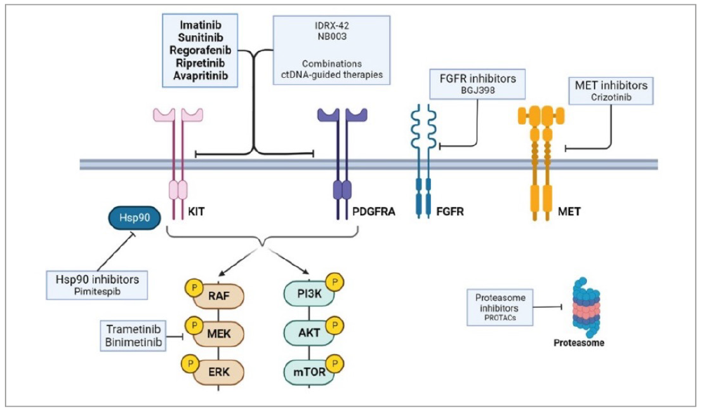
Among these, gastrointestinal stromal tumors (GISTs) represent the most common malignant mesenchymal neoplasm and constitutes a paradigm model for studying oncogene addiction and identifying structural and functional mechanisms of response and resistance to treatment. In recent years, our work has advanced the understanding of the pathophysiological mechanisms of GIST and exposed critical insights on the complex evolution of KIT/PDGFRA driver mutations leading to therapy resistance through comprehensive analyses of tissue and liquid biopsy samples. Currently ongoing efforts on GIST include: 1) The GISTomics project—a European initiative led by our group—aims to advance insights into the landscape of GIST evolution; and 2) A real-world-based genomic and transcriptomic landscape in a cohort of 1,427 GIST cases, the largest series available to date.
As part of our translational efforts, we pursue the identification of critical molecular mediators of therapeutic adaptation and resistance in GIST. Our work involves the discovery and functional characterization of key molecular events that can be potentially targetable. These mediators range from signaling intermediates, to the unfolded protein response (UPR) and interactions with the tissue microenvironment (TME), all of which are involved in the sensitivity/resistance to KIT inhibition. In this sense, César Serrano’s twin lab at the Institute for Research in Biomedicine (IRB), within the TRIP Clinics program, expands these views to the fields of protein degradation and the ubiquitin-proteasome system. Additionally, we leverage multi-omic datasets to identify and characterize the patterns and function of chromosome instability in sarcoma progression.
Our aim is to have a true clinical impact by improving the daily treatment and care of our patients. We are proud that our Sarcoma Multidisciplinary Unit is a Spanish Health Ministry-designed referral center (CSUR), and we belong to the European Reference Network ERN-EURACAN, which therefore constitutes an optimal setting for translating cancer discovery into clinical benefits. In this sense, three highlights from 2024 include the finalization of the MOTION trial, that confirmed the activity of vimseltinib in patients with tenosynovial giant cell tumor (TGCT) (Gelderblom et al, Lancet 2024); the launching of the phase III INSIGHT trial, which aim to validate in the clinic the use of ctDNA to guide therapeutic decisions in GIST (NCT05734105); the involvement in the updated guidelines from the European Society of Medical Oncology (ESMO) to establish the role of next-generation sequencing (NGS) in cancer, including sarcomas and other rare tumors.
- Identification of critical molecular mediators of oncogenic signaling in sarcomas.
- Studying the patterns of CIN in sarcomas together with its role in tumor evolution and progression.
- Characterization of mechanisms of response and resistance to targeted therapies in sarcomas, including preclinical modelling and validation of therapeutic strategies for their translation to the clinic.
- Phase I to phase III studies for clinical drug development in sarcomas.
- Our group leads cutting edge studies toward the clinical implementation of liquid biopsy in gastrointestinal stromal tumor (GIST).
- We have received critical support from several funding bodies to study the evolutionary landscape behind treatment resistance in GIST.
- César Serrano is the Secretary of the Spanish Society of Medical Oncology (SEOM) and the scientific coordinator of SEOM’s Annual Meeting.
- The support we receive from patients through the #SarcModel project enables us to generate preclinical models of most sarcoma subtypes.
- Identify novel patterns of sarcoma evolution and progression.
- Pursue ctDNA implementation in GIST to guide clinical decision making.
- Advance insights into the tumor microenvironment of sarcomas. These tumors are typically characterized by their ‘cold’ features.
- Preclinical and clinical drug development of novel compounds and/or combinations to transfer to the clinic.
- Heinrich MC, Jones RL, George S, Gelderblom H, Schöffski P, von Mehren M, Zalcberg JR, Kang YK, Razak AA, Trent J, Attia S, Le Cesne A, Siontis BL, Goldstein D, Boye K, Sanchez C, Steeghs N, Rutkowski P, Druta M, Serrano C, Somaiah N, Chi P, Reichmann W, Sprott K, Achour H, Sherman ML, Ruiz-Soto R, Blay JY, Bauer S. Ripretinib versus sunitinib in gastrointestinal stromal tumor: ctDNA biomarker analysis of the phase 3 INTRIGUE trial. Nat Med. 2024 Feb;30(2):498-506.
- Mühlenberg T, Falkenhorst J, Schulz T, Fletcher BS, Teuber A, Krzeciesa D, Klooster I, Lundberg M, Wilson L, Lategahn J, von Mehren M, Grunewald S, Tüns AI, Wardelmann E, Sicklick JK, Brahmi M, Serrano C, Schildhaus HU, Sievers S, Treckmann J, Heinrich MC, Raut CP, Ou WB, Marino-Enriquez A, George S, Rauh D, Fletcher JA, Bauer S. KIT ATP-Binding Pocket/Activation Loop Mutations in GI Stromal Tumor: Emerging Mechanisms of Kinase Inhibitor Escape. J Clin Oncol. 2024 Apr 20;42(12):1439-1449.
- Mosele MF, Westphalen CB, Stenzinger A, Barlesi F, Bayle A, Bièche I, Bonastre J, Castro E, Dienstmann R, Krämer A, Czarnecka AM, Meric-Bernstam F, Michiels S, Miller R, Normanno N, Reis-Filho J, Remon J, Robson M, Rouleau E, Scarpa A, Serrano C, Mateo J, André F. Recommendations for the use of next-generation sequencing (NGS) for patients with advanced cancer in 2024: a report from the ESMO Precision Medicine Working Group. Ann Oncol. 2024 Jul;35(7):588-606.
- Gelderblom H, Bhadri V, Stacchiotti S, Bauer S, Wagner AJ, van de Sande M, Bernthal NM, López Pousa A, Razak AA, Italiano A, Ahmed M, Le Cesne A, Tinoco G, Boye K, Martín-Broto J, Palmerini E, Tafuto S, Pratap S, Powers BC, Reichardt P, Casado Herráez A, Rutkowski P, Tait C, Zarins F, Harrow B, Sharma MG, Ruiz-Soto R, Sherman ML, Blay JY, Tap WD; MOTION investigators. Vimseltinib versus placebo for tenosynovial giant cell tumour (MOTION): a multicentre, randomised, double-blind, placebo-controlled, phase 3 trial. Lancet. 2024 Jun 22;403(10445):2709-2719.
- Spanish Society of Medical Oncology (SEOM), Scientific coordinator for SEOM 2024 and SEOM 20025 Annual Meetings.
- Determinantes clínicos y moleculares de respuesta y progresión a la inhibición de KIT en pacientes con tumores del estroma gastrointestinal (GIST) con respuesta prolongada a Imatinib. Grupo Español de Investigación en Sarcomas (GEIS), Sociedad Española de Oncología Médica (SEOM), and Fundación Mari Paz Jiménez Casado.
- Modulating our ubiquitination machinery to innovate treatments and drug discovery in cancer. Asociación Española Contra el Cáncer– AECC (Spanish Association against Cancer).
- Perfiles de resistencia molecular a la inhibición de KIT/PDGFRA en tumores del estroma gastrointestinal. Asociación Española Contra el Cáncer – AECC (Spanish Association against Cancer).
- Centralization of pathological diagnosis and implementation of precision medicine strategies in sarcomas. Asociación Española Contra el Cáncer – AECC (Spanish Association against Cancer).
- Genomic determinants of tumor evolution and progression in sarcomas with simple karyotype. CRIS Foundation.
- Drivers of chromosome instability in karyotypically simple sarcomas: gastrointestinal stromal tumor as a paradigm. Instituto de Salud Carlos III.
- Yin-yang modulation of the ubiquitin-proteasome system to chart and treat RTK-driven neoplasms. TRIP-Clinics program (La Caixa Foundation and Generalitat de Catalunya).
- Heinrich MC, Jones RL, George S, Gelderblom H, Schöffski P, von Mehren M, Zalcberg JR, Kang YK, Razak AA, Trent J, Attia S, Le Cesne A, Siontis BL, Goldstein D, Boye K, Sanchez C, Steeghs N, Rutkowski P, Druta M, Serrano C, Somaiah N, Chi P, Reichmann W, Sprott K, Achour H, Sherman ML, Ruiz-Soto R, Blay JY, Bauer S. Ripretinib versus sunitinib in gastrointestinal stromal tumor: ctDNA biomarker analysis of the phase 3 INTRIGUE trial. Nat Med. 2024 Feb;30(2):498-506.
- Mühlenberg T, Falkenhorst J, Schulz T, Fletcher BS, Teuber A, Krzeciesa D, Klooster I, Lundberg M, Wilson L, Lategahn J, von Mehren M, Grunewald S, Tüns AI, Wardelmann E, Sicklick JK, Brahmi M, Serrano C, Schildhaus HU, Sievers S, Treckmann J, Heinrich MC, Raut CP, Ou WB, Marino-Enriquez A, George S, Rauh D, Fletcher JA, Bauer S. KIT ATP-Binding Pocket/Activation Loop Mutations in GI Stromal Tumor: Emerging Mechanisms of Kinase Inhibitor Escape. J Clin Oncol. 2024 Apr 20;42(12):1439-1449.
- Mosele MF, Westphalen CB, Stenzinger A, Barlesi F, Bayle A, Bièche I, Bonastre J, Castro E, Dienstmann R, Krämer A, Czarnecka AM, Meric-Bernstam F, Michiels S, Miller R, Normanno N, Reis-Filho J, Remon J, Robson M, Rouleau E, Scarpa A, Serrano C, Mateo J, André F. Recommendations for the use of next-generation sequencing (NGS) for patients with advanced cancer in 2024: a report from the ESMO Precision Medicine Working Group. Ann Oncol. 2024 Jul;35(7):588-606.
- Gelderblom H, Bhadri V, Stacchiotti S, Bauer S, Wagner AJ, van de Sande M, Bernthal NM, López Pousa A, Razak AA, Italiano A, Ahmed M, Le Cesne A, Tinoco G, Boye K, Martín-Broto J, Palmerini E, Tafuto S, Pratap S, Powers BC, Reichardt P, Casado Herráez A, Rutkowski P, Tait C, Zarins F, Harrow B, Sharma MG, Ruiz-Soto R, Sherman ML, Blay JY, Tap WD; MOTION investigators. Vimseltinib versus placebo for tenosynovial giant cell tumour (MOTION): a multicentre, randomised, double-blind, placebo-controlled, phase 3 trial. Lancet. 2024 Jun 22;403(10445):2709-2719.
- George S, Blay JY, Chi P, Jones RL, Serrano C, Somaiah N, Gelderblom H, Zalcberg JR, Reichmann W, Sprott K, Cox P, Sherman ML, Ruiz-Soto R, Heinrich MC, Bauer S. The INSIGHT study: a randomized, Phase III study of ripretinib versus sunitinib for advanced gastrointestinal stromal tumor with KIT exon 11 + 17/18 mutations. Future Oncol. 2024;20(27):1973-1982. doi: 10.1080/14796694.2024.2376521. Epub 2024 Sep 4.
- Muñoz-Casares FC, Martín-Broto J, Cascales-Campos P, Torres-Melero J, López-Rojo I, Gómez-Barbadillo J, González-Bayón L, Sebio A, Serrano C, Carvalhal S, Abreu de Souza J, Souza A, Flores-Ayala G, Palacios Fuenmayor LJ, Lopes-Bras R, González-López JA, Vasques H, Asencio-Pascual JM. Ibero-American Consensus for the Management of Peritoneal Sarcomatosis: Updated Review and Clinical Recommendations. Cancers (Basel). 2024 Jul 25;16(15):2646. doi: 10.3390/cancers16152646.
- Carmona J, Chavarria E, Donoghue K, von Gertten C, Oberrauch P, Pailler E, Scoazec G, Weijer R, Balmaña J, Brana I, Brunelli C, Delaloge S, Deloger M, Delpy P, Ernberg I, Fitzgerald RC, Garralda E, Lablans M, Lëhtio J, Lopez C, Fernández M, Miceli R, Nuciforo P, Perez-Lopez R, Provenzano E, Schmidt MK, Serrano C, Steeghs N, Tamborero D, Wirta V, Baird RD, Barker K, Barlesi F, Baumann M, Bergh J, de Braud F, Fizazi K, Fröhling S, Piris-Giménez A, Seamon K, Van der Heijden MS, Zwart W, Tabernero J. Cancer Core Europe: Leveraging Institutional Synergies to Advance Oncology Research and Care Globally. Cancer Discov. 2024 Jul 1;14(7):1147-1153. doi: 10.1158/2159-8290.CD-24-0377.
- Gómez-Peregrina D, Cicala CM, Serrano C. Monitoring advanced gastrointestinal stromal tumor with circulating tumor DNA. Curr Opin Oncol. 2024 Jul 1;36(4):282-290. doi: 10.1097/CCO.0000000000001040. Epub 2024 Apr 19.
- Cicala CM, Olivares-Rivas I, Aguirre-Carrillo JA, Serrano C. KIT/PDGFRA inhibitors for the treatment of gastrointestinal stromal tumors: getting to the gist of the problem. Expert Opin Investig Drugs. 2024 Mar;33(3):159-170. doi: 10.1080/13543784.2024.2318317. Epub 2024 Feb 14.
- García-Díaz HC, Eremiev S, Gómez-Alonso J, Veas Rodriguez J, Farriols A, Carreras MJ, Serrano C. Hyperammonemic encephalopathy after tyrosine kinase inhibitors: A literature review and a case example. J Oncol Pharm Pract. 2024 Apr;30(3):576-583. doi: 10.1177/10781552231225188. Epub 2024 Jan 22.
- Serrano C, Rothschild S, Villacampa G, Heinrich MC, George S, Blay JY, Sicklick JK, Schwartz GK, Rastogi S, Jones RL, Rutkowski P, Somaiah N, Navarro V, Evans D, Trent JC. Novel trial designs for patients with gastrointestinal stromal tumor. ESMO Open. 2024 Jan;9(1):102218. doi: 10.1016/j.esmoop.2023.102218. Epub 2024 Jan 8.
- Heinrich MC, Zhang X, Jones RL, George S, Serrano C, Deng Y, Bauer S, Cai S, Wu X, Zhou Y, Tao K, Zheng Z, Zhang J, Cui Y, Cao H, Wang M, Hu J, Yang J, Li J, Shen L. Clinical Benefit of Avapritinib in KIT-Mutant Gastrointestinal Stromal Tumors: A Post Hoc Analysis of the Phase I NAVIGATOR and Phase I/II CS3007-001 Studies. Clin Cancer Res. 2024 Feb 16;30(4):719-728. doi: 10.1158/1078-0432.CCR-23-1861.
- Phase I, Open-label, Multicenter Study to Assess the Safety, Tolerability, and Pharmacokinetics of NB003 in Subjects with Advanced Malignancies. Role: PI; Steering Committee Member.
- StrateGIST: A First-in-human (FIH) Study of IDRX-42 in Participants With Metastatic and/or Unresectable Gastrointestinal Stromal Tumors (GIST). Role: PI; Advisory Board Member.
- INSIGHT: A Study of Ripretinib vs Sunitinib in Patients With Advanced GIST With Specific KIT Exon Mutations Who Were Previously Treated With Imatinib. Role: PI; Steering Committee Member.
- PEAK: A Phase III Randomized Trial of CGT9486+Sunitinib vs. Sunitinib in Subjects With Gastrointestinal Stromal Tumors. Role: PI; Advisory Board Member.
- MOTION: Study of Vimseltinib for Tenosynovial Giant Cell Tumor. Role: PI.
- VOYAGER: An International, Multicenter, Open-label, Randomized, Phase III Study of BLU-285 vs Regorafenib in Patients with Locally Advanced Unresectable or Metastatic Gastrointestinal Stromal Tumor (GIST). Role: National PI; Advisory Board Member
- INTRIGUE: A Phase III, Interventional, Randomized, Multicenter, Open-Label Study of DCC-2618 vs Sunitinib in Patients with Advanced Gastrointestinal Stromal Tumors after Treatment with Imatinib. Role: National PI; Advisory Board Member
- SELIGIST: A multicenter, phase Ib/II trial of Selinexor in combination with Imatinib in patients with metastatic and/or unresectable gastrointestinal stromal tumors (GISTs). Role: Global Trial Coordinator

Group Leader Maria Abad Research Assistant Lluis Palenzuela Graduate Students Alba Escriche, Marion Martinez
Cellular Plasticity and Cancer Group
We study the interplay between stress responses, cellular plasticity and cancer. Cellular plasticity is as a critical feature of cancer cells, enabling them to transit between different cellular states and promote tumor growth, disease progression after therapy, and metastasis.
We have previously reported that inducing cell dedifferentiation with the so-called Yamanaka factors can lead to the development of a variety of tumors. We have also demonstrated that tissue damage—the main driver of cancer—triggers the onset of cellular senescence which then induces dedifferentiation and the acquisition of stem cell properties in vivo. These findings have important therapeutic implications given that chemotherapy and radiotherapy—the cornerstones of treatment for most cancers—could have the side effect of inducing stemness in non-stem cancer cells and, in turn, possibly contribute to tumor recurrence and cancer cell spread.
Our main objective is to advance insights into the mechanisms and drivers implicated in this process, with the ultimate goal of developing novel therapies based on the inhibition of cancer cell plasticity.
Over the last few years, our efforts have mainly focused on identifying and characterizing novel cancer microproteins, encoded by previously assumed non-coding regions, which could be novel actors in carcinogenesis. We have discovered several new cancer microproteins and have obtained compelling evidence in vitro and in vivo that they act as novel tumor suppressors, inducing cell cycle arrest, differentiation or inhibition of mesenchymal traits in cancer cells.
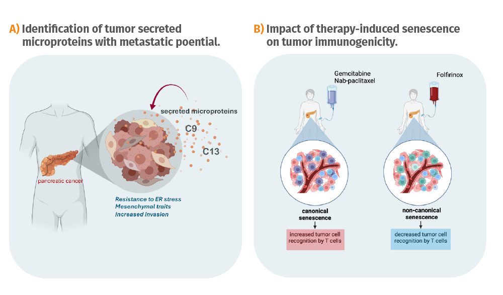
In recent years, using a proteogenomics approach, we have identified a set of microproteins secreted by pancreatic tumors, either soluble or secreted in exosomes. Importantly, many of these microproteins are also detected in patients’ plasma. In 2024 we have deeply characterized two of them, C9 and C13, and demonstrated that they have a pro-metastatic role in preclinical mouse models, modulating the unfolded-protein response and inducing mesenchymal treats in cancer cells. Our results suggest that microproteins are crucial cellular messengers for pancreatic cancer metastasis and they could also serve as novel cancer biomarkers for the early detection of disease and patient stratification for tailored therapies, as well as therapeutic targets.
We have also continued working on our collaborative project to study therapy-induced tumoral senescence and its potential as a therapeutic target, funded by the Asociación Española Contra el Cáncer - AECC (Spanish Association Against Cancer). Notably, we have observed that different chemotherapies currently used in the clinic to treat pancreatic cancer induce different types of cellular senescence, which intriguingly have opposite effects on the recognition of tumor cells by the tumor infiltrating lymphocytes (TILs).
Some drugs induce a type of senescence that increases tumor cell recognition by TILs, while others impair tumor cell recognition. Importantly, the ability of some drugs to induce tumor recognition correlates with their potential to activate the IFNg pathway and the antigen presenting machinery. These are very interesting results with obvious clinical implications that my guide the development of futured tailored therapies.
- Advance insights into the interplay between therapy-induced senescence, cellular plasticity and cancer.
- Decipher the molecular mechanisms governing the acquisition of stem cell properties during tumorigenesis and after therapy.
- Discover and characterize novel microproteins involved in cancer cell plasticity.
- Develop novel anti-cancer therapies based on the inhibition of cancer cell plasticity.
- Marion Martinez defended her doctoral thesis: Defining the cecreted microproteome for novel regulators of intercellular communication in pancreatic cancer, and was awarded Cum Laude distinction.
- We have shown that C9 and C13 microproteins promote pancreatic cancer metastasis by modulating the unfolded protein response (UPR) and inducing epithelial-to-mesenchymal (EMT) transition in cancer cells.
- We have demonstrated that different chemotherapies currently used in the clinic to treat pancreatic cancer induce different types of cellular senescence, which intriguingly have opposite effects on the recognition of tumor cells by tumor infiltrating lymphocytes (TILs).
- In December 2024, our group moved from VHIO to the Cambridge Institute of Science, Altos Labs, Cambridge, UK.
- Shen YR, Zaballa S, Bech X, Sancho-Balsells A, Rodríguez-Navarro I, Cifuentes-Díaz C, Seyit-Bremer G, Chun SH, Straub T, Abante J, Merino-Valverde I, Richart L, Gupta V, Li HY, Ballasch I, Alcázar N, Alberch J, Canals JM, Abad M, Serrano M, Klein R, Giralt A, Del Toro D. Expansion of the neocortex and protection from neurodegeneration by in vivo transient reprogramming. Cell Stem Cell. 2024 Dec 5;31(12):1741-1759.e8.
- Grantor: Spanish Association Against Cancer - AECC. Title: Tumoral senescence induced by anti-cancer therapies constitutes a novel prognostic biomarker and a therapeutic target. Reference: PRYCO211023SERR. Duration: 9/2021-8/2026. Co-PI: Maria Abad.

Group Leader Rodrigo A. Toledo PhD Students Carlota Arenillas, Oriol Mirallas Viñas, Ana Belen Moreno-Cárdenas Data Manager Alma Calahorro Laboratory Technician Aya Louhamane Bioinformaticians Adriana Bareas, Alessandra Bonilla, Debayan Datta Data Scientist Kira Raskina
Biomarkers and Clonal Dynamics Group
VHIO’s Biomarkers and Clonal Dynamics Group applies innovative tumor evolution analyses on longitudinal tumor and circulating tumor DNA (ctDNA) liquid biopsy samples across diverse clinical and therapeutic contexts. Through this integrative approach, our research aims at unravelling the evolutionary complexity of human cancers and expose actionable insights that may be translated into clinical practice, with a primary focus on cancer immunotherapy and targeted therapies.
Our liquid biopsy research also extends to rare adrenal malignancies, including pheochromocytomas and paragangliomas (PPGL) as well as adrenocortical carcinomas (ACC), where we apply ctDNA-based approaches to improve disease monitoring and therapeutic decision-making.
- Apply multimodal ctDNA assays to predict response to immunotherapy and targeted therapies across tumor types.
- Characterize tumor heterogeneity: examine the temporal heterogeneity of tumors using tumor and liquid biopsy sequencing approaches during treatment response and disease progression, particularly in the context of immunotherapy and targeted therapies.
- Investigate the mechanisms of cancer drug resistance to immunotherapy and targeted therapies to identify potential biomarkers of resistance in liquid biopsy that inform more effective treatment strategies.
- Evaluate the clinical utility of circulating tumor DNA (ctDNA) analysis in rare adrenal cancers suh as pheochromocytomas, paragangliomas (PPGL), and adrenocortical carcinomas (ACC).
The IMMUNOMICs Liquid Biopsy Project
Our laboratory has developed a comprehensive liquid biopsy repository, comprising longitudinal tumor and plasma samples from cancer patients undergoing immunotherapy. The IMMUNOMICs cohort, which includes >500 patients with >4,000 longitudinal timepoints, serves as the foundation for applying multimodal liquid biopsy analyses, including:
- Ultrasensitive tumor-genome ctDNA profiling
- Whole-exome sequencing (WES) of cell-free DNA (cfDNA)
- Shallow whole-genome sequencing (sWGS)
This project aims to provide novel insights into the utility of different liquid biopsy approaches in characterizing baseline and early dynamic ctDNA profiles in immunotherapy-treated patients. We will also correlate these findings with treatment response and clinical outcomes, improving our ability to monitor and predict therapeutic efficacy.
The Rare Cancer Liquid Biopsy Project
In collaboration with the European Network for Adrenal Tumors (ENSAT), the A5 Adrenal Alliance, and the European Liquid Biopsy Society (ELBS), we have established a Global Consortium for Liquid Biopsy in Rare Adrenal Tumors. This initiative aims to advance the application of liquid biopsy in PPGL and ACC for enabling biomarker discovery, improving disease monitoring, and guiding therapeutic decision-making.
- IMMUNOMICs – Liquid biopsy analysis in cancer patients treated with immunotherapy.
- PREDICT – Liquid biopsy-based prediction of clinical outcomes in cancer immunotherapy.
- mPPGL – Circulating tumor DNA (ctDNA) as a prognostic biomarker in rare adrenal tumors.
- HYPOXIA – Positive selection of EPAS1-HIF2α mutations in systemic hypoxia.
- Wyatt, A. W., Litiere, S., Bidard, F. C., Cabel, L., Dyrskjøt, L., Karlovich, C. A., Pantel, K., Petrie, J., Philip, R., Andrews, H. S., Vellanki, P. J., Tolmeijer, S. H., Villalobos Alberu, X., Alfano, C., Bogaerts, J., Calvo, E., Chen, A. P., Toledo, R. A., de Vries, E. G. E., … RECIST ctDNA workshop group. (2024). Plasma ctDNA as a treatment response biomarker in metastatic cancers: Evaluation by the RECIST working group. Clinical Cancer Research, 30(22), 5034-5041. https://doi.org/10.1158/1078-0432.CCR-24-1883
- Stanley, K. E., Jatsenko, T., Tuveri, S., Sudhakaran, D., Lannoo, L., Van Calsteren, K., de Borre, M., Van Parijs, I., Van Coillie, L., Van Den Bogaert, K., De Almeida Toledo, R., Lenaerts, L., Tejpar, S., Punie, K., Rengifo, L. Y., Vandenberghe, P., Thienpont, B., & Vermeesch, J. R. (2024). Cell type signatures in cell-free DNA fragmentation profiles reveal disease biology. Nature Communications, 15(1), 2220. https://doi.org/10.1038/s41467-024-46435-0
- Prior, O., Macarro, C., Navarro, V., Monreal, C., Ligero, M., Garcia-Ruiz, A., Serna, G., Simonetti, S., Braña, I., Vieito, M., Escobar, M., Capdevila, J., Byrne, A. T., Dienstmann, R., Toledo, R., Nuciforo, P., Garralda, E., Grussu, F., Bernatowicz, K., & Perez-Lopez, R. (2024). Identification of precise 3D CT radiomics for habitat computation by machine learning in cancer. Radiology: Artificial Intelligence, 6(2), e230118. https://doi.org/10.1148/ryai.230118
- Ligero, M., Serna, G., El Nahhas, O. S. M., Sansano, I., Mauchanski, S., Viaplana, C., Calderaro, J., Toledo, R. A., Dienstmann, R., Vanguri, R. S., Sauter, J. L., Sanchez-Vega, F., Shah, S. P., Ramón Y Cajal, S., Garralda, E., Nuciforo, P., Perez-Lopez, R., & Kather, J. N. (2024). Weakly supervised deep learning predicts immunotherapy response in solid tumors based on PD-L1 expression. Cancer Research Communications, 4(1), 92-102. https://doi.org/10.1158/2767-9764.CRC-23-0287

Group Leader Laura Soucek Senior Investigators Jonathan R. Whitfield, Mariano F. Zacarías-Fluck Postdoctoral Fellow Ana Lopez Garza Technicians Judit Grueso, Romina Mariel Rodriguez, Melisa Morcillo Sanchez, Erika Serrano del Pozo, Manrique Jose Valdez-Bango Martín PhD Students Daniel Capitán Leo, Fabio Giuntini, Íñigo González-Larreategui Visiting Student Mélodie Oullet Master's Student Manuel Lillo
Models of Cancer Therapies Group
Our group focuses on the pleiotropic and ubiquitous MYC oncoprotein, whose deregulation is implicated in almost all human cancers. The technical challenges of targeting nuclear transcription factors such as MYC –and the concern regarding potential side effects–had until recently precluded any clinical validation of MYC inhibition as a possible therapeutic strategy.
Over the past years, we have demonstrated in several mouse models that MYC inhibition has a dramatic therapeutic impact across several tumor types, with very mild and reversible side effects in normal tissue. Encouraged by our results in mice, we created a spin-off company, Peptomyc S.L., for the development of MYC-inhibiting peptides for cancer therapy. In partnership with Peptomyc, our laboratory is currently validating our novel approach against difficult-to-treat cancers that are in dire need of new therapeutic options.
In 2024 we reported the results of a phase I clinical trial with the first Omomyc-derived compound, OMO-103. This compound is the first direct MYC inhibitor to successfully advance into clinical development. Building on these promising results, a phase Ib trial is currently ongoing in metastatic pancreatic ductal adenocarcinoma patients in combination with standard of care. The study is sponsored by Peptomyc and performed in 6 Spanish hospitals including the Vall d’Hebron University Hospital – HUVH.
We have continued to contribute to cancer research in general and, more specifically as a leading group in the MYC field, by authoring articles and reviews exploring new aspects of MYC biology in different pathologies and showcasing our efforts in developing a clinically viable MYC inhibitor.
- Preclinically validate novel therapeutic strategies against MYC in breast, brain, lung, neuroblastoma, melanoma, ovarian, and colorectal cancer.
- Validate Omomyc-based cell-penetrating mini-proteins against MYC for cancer therapy.
- Identify and validate biomarkers of response to MYC inhibition.
- Define the role of MYC in promoting cancer immune evasion and test the efficacy of MYC inhibitors and IO combination therapies in cancer treatment.
- Develop potent combinations of Omomyc with current therapeutics, including PARP inhibitors (PARPi) in the context of DNA Damage Response (DDR) deficient tumors, and KRAS inhibitors (KRASi).
- Investigate how the MYC network functions in MAX-defective gastrointestinal stromal tumors (GISTs) and small-cell lung cancer (SCLC) to define actionable targets to tackle these unmet clinical needs.
- The results from the phase I trial of OMO-103 were published in Nature Medicine with Laura Soucek as corresponding author, alongside Elena Garralda, Director of VHIO’s Research Unit for Molecular Therapy of Cancer (UITM) – CaixaResearch, and several members of the lab and Peptomyc.
- Two academic collaborations led to 2 research papers: one with Gianluca and Arturo Sala in the Journal of Controlled Release describing the efficacy of an Omomyc antibody-drug conjugate for specific targeting of HER3-positive tumours, the other with Joaquin Arribas to develop HER2-targeting CAR-T cells.
- Reflective of Laura Soucek’s role as a key opinion leader in the field, other publications this year include reviews in Signal Transduction and Targeted Therapy and GEN Biotechnology.
- After assembling a Special Collection for Frontiers in Cell and Developmental Biology on the potential of MYC as a therapeutic target beyond cancer, Senior Researchers in the lab Jonathan R. Whitfield and Mariano F. Zacarías-Fluck, along with Laura Soucek, wrote a review on the topic. Jonathan Whitfield also wrote the Editorial for this Special Collection.
- Daniel Capitán Leo was awarded with the prestigious predoctoral ”la Caixa” predoctoral INPhINIT Retaining fellowship.
- Fabio Giuntini successfully applied to the CAIMI/BBVA Secondment Programme of VHIO to perform a 3-month placement at University of Cambridge. He also gained an EACR Travel Grant, along with PhD student Íñigo González-Larreategui.
- Íñigo González-Larreategui was also selected to give an oral presentation at the XVIII Congress of the Catalan Society of Immunology (SCI), and Fabio Giuntini won a poster prize at the Karolinska Istitutet MYC Meeting.
- We have been pleased to host in the lab Mélodie Oullet, a visiting student from Canada, under the prestigious Mitacs program.
- Advance the validation of novel therapies for cancer treatment, in particular against the previously undruggable target MYC.
- Define the role of MYC in promoting cancer immune evasion and assess the potential of MYC inhibition in combination with immune-oncology therapies and personalized medicine.
- Explore the role of the MYC network members in MAX-null tumors and identify potentially actionable targets and therapeutic strategies against these cancers.
- Garralda E, Beaulieu ME, Moreno V, Casacuberta-Serra S, Martínez-Martín S, Foradada L, Alonso G, Massó-Vallés D, López-Estévez S, Jauset T, Corral de la Fuente E, Doger B, Hernández T, Perez-Lopez R, Arqués O, Castillo Cano V, Morales J, Whitfield JR, Niewel M, Soucek L, Calvo E. MYC targeting by OMO-103 in solid tumors: a phase 1 trial. Nat Med. 2024 Mar;30(3):762-771.
- Román Alonso M, Grinyó-Escuer A, Duro-Sánchez S, Rius-Ruiz I, Bort-Brusca M, Escorihuela M, Maqueda-Marcos S, Pérez-Ramos S, Gago J, Nogales V, Espinosa-Bravo M, Peg V, Escrivá-de-Romaní S, Foradada L, Soucek L, Braña I, Galvao V, Martín-Lluesma S, Moessner E, Klein C, Garralda E, Saura C, Arribas J. Generation of chimeric antigen receptor T cells targeting p95HER2 in solid tumors. Nat Commun. 2024 Nov 18;15(1):9589.
- Casacuberta-Serra S, González-Larreategui Í, Capitán-Leo D, Soucek L. MYC and KRAS cooperation: from historical challenges to therapeutic opportunities in cancer. Signal Transduct Target Ther. 2024 Aug 21;9(1):205.
- Zacarías-Fluck MF, Soucek L, Whitfield JR. MYC: there is more to it than cancer. Front Cell Dev Biol. 2024 Mar 6;12:1342872.
- Laura Soucek was awarded with an ERC Advanced Grant, with 2.2 M € to fund the next 5 years’ research on optimizing the clinical applicability of the MYC inhibitor Omomyc and further understanding MYC biology in DNA damage response and MAX-null tumors.
- Daniel Capitán Leo was successful in 3 PhD grant applications, and was awarded a with a prestigious predoctoral "La Caixa" predoctoral INPhINIT Retaining fellowship.
- Fabio Giuntini successfully applied to the CAIMI/BBVA Secondment Program of VHIO for a 3-month placement at the University of Cambridge, UK. He was also awarded with an EACR Travel Grant, along with Íñigo González-Larreategui, a fellow VHIO PhD student.
- A new patent application was submitted (Patent No. EP24382257.4) for "COMPOUND FOR THE TREATMENT OF AUTOIMMUNE DISEASES”. Inventors: Sílvia Casacuberta-Serra, Marie-Eve Beaulieu, Laura Soucek, Carmen Espejo Ruiz ,and Herena Eixarch Ahufinger.
- Laura Soucek was selected to appear on the Advent Calendar showcased on the front of Casa Seat building in central Barcelona. The images of the 24 featured Catalonia-based luminaries across different professions were drawn by the famous neo-pop street artist TVBoy.
- Chiara Giorgetti Award from the Spanish Association of Metastatic Breast Cancer.
Title: Combining MYC and PARP inhibitors as a novel therapeutic strategy against triple-negative breast cancer.
Duration: 2022-2024.
PI: Laura Soucek. - 2nd BBVA Foundation Comprehensive Program of Cancer Immunotherapy & Immunology (CAIMI II), VHIO-FBBVA Collaboration.
Project titles: Understanding the tumor microenvironment to identify therapeutic targets and improve the response to immune checkpoint inhibitors.
Understanding the mechanisms of resistance to IT and immune evasion to targeted therapies and designing new rational combinatory treatments. Duration: 2022-2025.
PI: Laura Soucek. - Grant: I+D+I en Líneas Estratégicas en colaboración público-privada from the Spanish Ministry of Economy, Industry and Competitiveness.
Title: Using MYC inhibition to overcome immunotherapy resistance in KRAS-driven NSCLC with diverse mutational profiles (MYCOMBIO).
Duration: 2021-2024.
PI: Laura Soucek. - Grant: Proyectos en Colaboración Público-Privado from the Spanish Ministry of Sciences, Innovation and Universities.
Title: Clinical proof-of-concept of a first-in-class MYC inhibitor in combination therapy and expansion of its development plan (CLINIMYC).
Duration: 2023-2026.
PI: Laura Soucek. - AECC 70% Survivorship
Title: Small cell lung cancer: from biology networks to tailored therapy (SOSCLC)
Duration: 2024-2029
Co-applicant: Laura Soucek - Grant: ERC Advanced Grant
Title: Maximizing the use of a first clinically viable MYC inhibitor (MYCiMAX)
Duration: 2024-2029
PI: Laura Soucek
- Garralda E, Beaulieu ME, Moreno V, Casacuberta-Serra S, Martínez-Martín S, Foradada L, Alonso G, Massó-Vallés D, López-Estévez S, Jauset T, Corral de la Fuente E, Doger B, Hernández T, Perez-Lopez R, Arqués O, Castillo Cano V, Morales J, Whitfield JR, Niewel M, Soucek L*, Calvo E. MYC targeting by OMO-103 in solid tumors: a phase 1 trial. Nat Med. 2024 Mar;30(3):762-771. doi: 10.1038/s41591-024-02805-1. Epub 2024 Feb 6.
*Corresponding author. - Román Alonso M, Grinyó-Escuer A, Duro-Sánchez S, Rius-Ruiz I, Bort-Brusca M, Escorihuela M, Maqueda-Marcos S, Pérez-Ramos S, Gago J, Nogales V, Espinosa-Bravo M, Peg V, Escrivá-de-Romaní S, Foradada L, Soucek L, Braña I, Galvao V, Martín-Lluesma S, Moessner E, Klein C, Garralda E, Saura C, Arribas J. Generation of chimeric antigen receptor T cells targeting p95HER2 in solid tumors. Nat Commun. 2024 Nov 18;15(1):9589. doi: 10.1038/s41467-024-53265-7.
- Casacuberta-Serra S, González-Larreategui Í, Capitán-Leo D, Soucek L. MYC and KRAS cooperation: from historical challenges to therapeutic opportunities in cancer. Signal Transduct Target Ther. 2024 Aug 21;9(1):205. doi: 10.1038/s41392-024-01907-z.
- Zacarías-Fluck MF, Soucek L*, Whitfield JR. MYC: there is more to it than cancer. Front Cell Dev Biol. 2024 Mar 6;12:1342872. doi: 10.3389/fcell.2024.1342872.
*Corresponding author. - Román Alonso M, Grinyó-Escuer A, Duro-Sánchez S, Rius-Ruiz I, Bort-Brusca M, Escorihuela M, Maqueda-Marcos S, Pérez-Ramos S, Gago J, Nogales V, Espinosa-Bravo M, Peg V, Escrivá-de-Romaní S, Foradada L, Soucek L, Braña I, Galvao V, Martín-Lluesma S, Moessner E, Klein C, Garralda E, Saura C, Arribas J. Generation of chimeric antigen receptor T cells targeting p95HER2 in solid tumors. Nat Commun. 2024 Nov 18;15(1):9589. doi: 10.1038/s41467-024-53265-7.
- Bibbò S, Capone E, Lovato G, Ponziani S, Lamolinara A, Iezzi M, Lattanzio R, Mazzocco K, Morini M, Giansanti F, De Laurenzi V, Whitfield J, Iacobelli S, Ippoliti R, Beaulieu ME, Soucek L, Sala A, Sala G. EV20/Omomyc: A novel dual MYC/HER3 targeting immunoconjugate. J Control Release. 2024 Oct;374:171-180. doi: 10.1016/j.jconrel.2024.08.009. Epub 2024 Aug 14.
- Hugo Thabussot, Marie-Beaulieu and Laura Soucek. Stretching the Limits of Bicyclic Peptides: A Novel Approach to MYC Inhibition and Beyond. GEN Biotechnology. Volume 3, Number 1, 2024 Feb 15. https://doi.org/10.1089/genbio.2024.0003. News and Views.
- Whitfield JR. Editorial: MYC as a disease target beyond cancer. Front Cell Dev Biol. 2024 Aug 22;12:1467372. doi: 10.3389/fcell.2024.1467372.
- Phase 1b with OMO-103 in combination with standard of care chemotherapy in first line metastatic pancreatic ductal adenocarcinoma patients (NCT06059001).

Group Leader Violeta Serra Postdoctoral Fellows Andrea Herencia Alba Llop-Guevara, Laia Monserrat, Flaminia Pedretti Graduate Students Heura Domènech, Andreu Òdena Visiting Students Ruth Maldonado, Taras Yuviz Technicians Marta Guzmán, Cristina Molina, Olga Rodríguez
Experimental Therapeutics Group
We conduct bench-to-bedside preclinical research in breast cancer to advance insights into biomarkers of response to targeted therapies. To do so, we generate preclinical models including patient-derived xenografts (PDXs) and patient-derived cultures (PDCs) from primary or metastatic breast cancer.
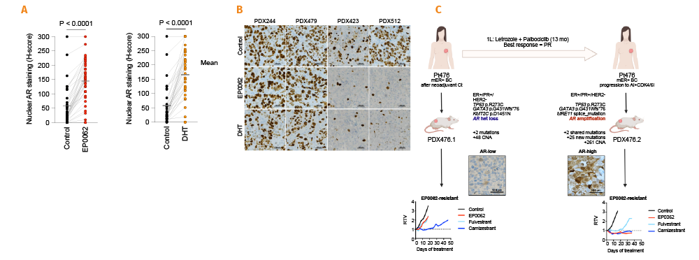
We have contributed to the field of endocrine resistance and continue to advance insights into mechanisms of sensitivity and resistance to CDK4/6 inhibitors, FGFR inhibitors, AKT inhibitors, and AR modulators (SARMs) in breast tumors. Using clinically relevant PDXs, we have provided evidence that the loss of G1-cell cycle checkpoint control, such as mutation/loss of RB1 or CCND1- amplification, is associated with the lack of response to CDK4/6 blockade in estrogen receptor-positive (ER+) breast cancer. Additionally, we have generated a collection of ER+ PDXs for testing novel therapies and treatment combinations in this setting.
We are also currently exploring the potential of a novel antibody-drug conjugate (ADC) as a therapeutic strategy for advanced breast cancers that have acquired resistance to standard of care treatments.
Encouraged by the early success of DNA damage repair inhibitors in patients with BRCA1/2-associated cancers, we initiated a project to identify biomarkers of response to PARP inhibitors (PARPi) as well as other DNA damage repair inhibitors including those targeting WEE1, ATR or POLQ.
Our studies underpin the capacity of tumors harboring BRCA1/2 mutations to recover DNA repair functionality and develop resistance to PARPi. We have developed an assay, the RAD51 test, which accurately identifies BRCA1/2-associated tumors that have restored DNA repair functionality and become resistant to these therapies. Importantly, this test also identifies tumors that are sensitive to PARPi through alterations in DNA repair by homologous recombination beyond the germline BRCA1/2 condition. We filed a patent (EU application in 2017 and PCT in 2018), and we are currently validating the use of this test in tumor samples from breast, ovarian, pancreatic, and prostate cancer patients.
Finally, we have also investigated the effects of PARPi on the tumor immune environment. DNA repair-deficient tumors accumulate cytosolic DNA, which can elicit an innate immune signal (the STING pathway) and upregulate interferon-related genes. Moreover, cGAS-STING-IFN signaling can be harnessed by combining PARP inhibitors with STING agonists to overcome acquired resistance and requires natural killer (NK) cells to mediate antitumor immunity.
Our group works closely together with Cristina Saura’s Breast Cancer Group, and Judith Balmaña’s Hereditary Cancer Genetics Group. Reflective of VHIO’s purely multidisciplinary and translational approach, our research is also carried out in collaboration with other groups including VHIO’s Molecular Oncology Group, Cancer Genomics Group, and Oncology Data Science – ODysSey Group, directed by Paolo Nuciforo, Ana Vivancos and Rodrigo Dienstmann, respectively.
In short, our group has significantly advanced insights into the mode of action of novel targeted therapies, identified new response biomarkers, and developed a biomarker-based assay with potential clinical application. We have also demonstrated the efficacy of hypothesis-based drug combinations.
- Develop predictive biomarkers of targeted treatments in estrogen receptor-positive (ER+) breast cancer and triple-negative breast cancer (TNBC), including inhibitors directed against DNA damage response (PARP, ATR, POLQ) as well as signaling/cell cycle kinases (SARM, CDK4/6, cyclin A/B).
- Explore novel treatment combinations for ER+ breast cancer and TNBC.
- Contribute to personalized medicine by developing diagnostic tests to better guide treatment strategies based on targeted therapies.
- Establish patient tumor-derived breast cancer preclinical models to explore hypothesis-based combinatorial therapies.
- Our group has advanced insights into the underlying mechanisms of resistance to poly ADP ribose polymerase inhibitors (PARPi) in breast cancer.
- We have demonstrated the potent and durable antitumor activity of a novel PARP1-selective inhibitor in patient-derived BRCA1/2-associated cancer models, which is currently in clinical development.
- We have shown that the prevalence of homologous recombination deficiency (HRD) in germline RAD51C/D breast and ovarian cancers is lower than in BRCA1/BRCA2 or PALB2-mutated tumors.
- We have established a panel of over one hundred ER+ breast cancer and TNBC patient-derived xenografts (PDXs), mainly from the metastatic setting. We focus on models that recapitulate progression on CDK4/6 inhibitors, and BRCA1/2-associated tumors.
- Validation of the homologous repair deficiency (HRD) biomarker based on the detection of RAD51 in retrospective and prospective clinical trials.
- Optimization of the RAD51 assay to be used in liquid biopsies (CTCs in blood) and automatization of the staining and image analysis of the test to facilitate its future implementation in the clinic.
- Identification of mechanisms of resistance to CDK4/6 inhibitors and the study of novel therapeutic strategies including AR modulators or antibody-drug conjugates (ADCs) for the treatment of advanced breast cancer. Based on these results, new patient stratification and therapeutic algorithms will be proposed for improving outcomes in patients who do not derive clinical benefit from current therapies.
- Unveil genetic and epigenetic mechanisms of acquired resistance to PARP inhibitors (PARPi) in hereditary BRCA1/2 breast cancer and beyond; by other HRR alterations. Propose novel therapeutic strategies for patients who do not respond to these drugs.
- Dissect the role of the STING pathway in BRCA mutated tumors and explore the combination of PARPi and therapies exploiting the autologous immune response.
- Continue expanding the panel of patient tumor-derived breast cancer models to investigate hypothesis-based, clinically applicable therapy combinations in breast cancer aimed at overcoming resistance to both anti-estrogen/CDK4/6 therapy and PARPi.
- Herencia-Ropero A, Llop-Guevara A, Staniszewska AD, Domènech-Vivó J, García-Galea E, Moles-Fernández A, Pedretti F, Domènech H, Rodríguez O, Guzmán M, Arenas EJ, Verdaguer H, Calero-Nieto FJ, Talbot S, Tobalina L, Leo E, Lau A, Nuciforo P, Dienstmann R, Macarulla T, Arribas J, Díez O, Gutiérrez-Enríquez S, Forment JV, O'Connor MJ, Albertella M, Balmaña J, Serra V. The PARP1 selective inhibitor saruparib (AZD5305) elicits potent and durable antitumor activity in patient-derived BRCA1/2-associated cancer models. Genome Med. 2024 Aug 26;16(1):107.
- Brasó-Maristany F, Ferrero-Cafiero JM, Falato C, Martínez-Sáez O, Cejalvo JM, Margelí M, Tolosa P, Salvador-Bofill FJ, Cruz J, González-Farré B, Sanfeliu E, Òdena A, Serra V, Pardo F, Luna Barrera AM, Arumi M, Guerra JA, Villacampa G, Sánchez-Bayona R, Ciruelos E, Espinosa-Bravo M, Izarzugaza Y, Galván P, Matito J, Pernas S, Vidal M, Santhanagopal A, Sellami D, Esker S, Fan PD, Suto F, Vivancos A, Pascual T, Prat A, Oliveira M. Patritumab deruxtecan in HER2-negative breast cancer: part B results of the window-of-opportunity SOLTI-1805 TOT-HER3 trial and biological determinants of early response. Nat Commun. 2024 Jul 11;15(1):5826.
- Torres-Esquius S, Llop-Guevara A, Gutiérrez-Enríquez S, Romey M, Teulé À, Llort G, Herrero A, Sánchez-Henarejos P, Vallmajó A, González-Santiago S, Chirivella I, Cano JM, Graña B, Simonetti S, Díaz de Corcuera I, Ramon Y Cajal T, Sanz J, Serrano S, Otero A, Churruca C, Sánchez-Heras AB, Servitja S, Guillén-Ponce C, Brunet J, Denkert C, Serra V, Balmaña J. Prevalence of Homologous Recombination Deficiency Among Patients With Germline RAD51C/D Breast or Ovarian Cancer. JAMA Netw Open. 2024 Apr 1;7(4):e247811.
- Harvey-Jones E, Raghunandan M, Robbez-Masson L, Magraner-Pardo L, Alaguthurai T, Yablonovitch A, Yen J, Xiao H, Brough R, Frankum J, Song F, Yeung J, Savy T, Gulati A, Alexander J, Kemp H, Starling C, Konde A, Marlow R, Cheang M, Proszek P, Hubank M, Cai M, Trendell J, Lu R, Liccardo R, Ravindran N, Llop-Guevara A, Rodriguez O, Balmana J, Lukashchuk N, Dorschner M, Drusbosky L, Roxanis I, Serra V, Haider S, Pettitt SJ, Lord CJ, Tutt ANJ. Longitudinal profiling identifies co-occurring BRCA1/2 reversions, TP53BP1, RIF1 and PAXIP1 mutations in PARP inhibitor-resistant advanced breast cancer. Ann Oncol. 2024 Apr;35(4):364-380.
- 2024-2027: Member of the Pelotonia-American Association for Cancer Research (AACR) Women’s Cancers Initiative Scientific Advisory Committee.
- 2024: Member of the Small Molecule Therapeutic Agents Section of the AACR Annual Meeting 2024 Program Committee.
- Title: Effect of Cyclin RxL inhibitors in PDXs of RB1-dysregulated and/or E2F pathway-altered breast cancer. Principal Investigator: Violeta Serra. Funding: Cycle Pharma. Duration: 2024-2025. Ongoing projects
- Title: Utilidad clínica de testar RAD51 como test de DIAgNósTico de HRD funcional para cáncer de mama (RADIANT), (PI23/00506). Principal Investigator: Violeta Serra. Funding: ISCIII-Project Grant. Duration: 2024-2026.
- Title: Identification of response biomarkers of a novel HER3-topoisomerase I inhibitor antibody drug conjugate (U3-1402) using breast cancer patient-derived xenoimplant models. Principal Investigators: Violeta Serra. Funding: Metastatic Breast Cancer Association. Duration: 2022-2024.
- Title: Development of new therapeutic strategies to overcome resistance to CDK4/6 inhibitors in breast cancers with expression of the estrogen receptor” (PI20/00892). Principal Investigators: Violeta Serra and Meritxell Bellet. Funding: ISCIII-Project Grant. Duration: 2021-2024.
- Title: Development of a liquid biopsy test to assess the homologous recombination function status in BRCA1/2 mutation carriers with breast cancer to inform therapy selection. Principal Investigators: Violeta Serra and Judith Balmaña. Funding: FERO Foundation Duration: 2019-2024.
- Title: Acknowledged Consolidated Research Group (SGR): Experimental Therapeutics in Breast Cancer (2021 SGR 01510). Coordinating Group: Violeta Serra. Funding: AGAUR-SGR. Duration: 2022-2024.
- Voulgarelis D, Forment JV, Herencia Ropero A, Polychronopoulos D, Cohen-Setton J, Bender A, Serra V, O'Connor MJ, Yates JWT, Bulusu KC. Understanding tumour growth variability in breast cancer xenograft models identifies PARP inhibition resistance biomarkers. NPJ Precis Oncol. 2024 Nov 18;8(1):266. doi: 10.1038/s41698-024-00702-x.
- Glaviano A, Wander SA, Baird RD, Yap KC, Lam HY, Toi M, Carbone D, Geoerger B, Serra V, Jones RH, Ngeow J, Toska E, Stebbing J, Crasta K, Finn RS, Diana P, Vuina K, de Bruin RAM, Surana U, Bardia A, Kumar AP. Mechanisms of sensitivity and resistance to CDK4/CDK6 inhibitors in hormone receptor-positive breast cancer treatment. Drug Resist Updat. 2024 Sep;76:101103. doi: 10.1016/j.drup.2024.101103. Epub 2024 Jun 25.
- Herencia-Ropero A, Llop-Guevara A, Staniszewska AD, Domènech-Vivó J, García-Galea E, Moles-Fernández A, Pedretti F, Domènech H, Rodríguez O, Guzmán M, Arenas EJ, Verdaguer H, Calero-Nieto FJ, Talbot S, Tobalina L, Leo E, Lau A, Nuciforo P, Dienstmann R, Macarulla T, Arribas J, Díez O, Gutiérrez-Enríquez S, Forment JV, O'Connor MJ, Albertella M, Balmaña J, Serra V. The PARP1 selective inhibitor saruparib (AZD5305) elicits potent and durable antitumor activity in patient-derived BRCA1/2-associated cancer models. Genome Med. 2024 Aug 26;16(1):107. doi: 10.1186/s13073-024-01370-z.
- Brasó-Maristany F, Ferrero-Cafiero JM, Falato C, Martínez-Sáez O, Cejalvo JM, Margelí M, Tolosa P, Salvador-Bofill FJ, Cruz J, González-Farré B, Sanfeliu E, Òdena A, Serra V, Pardo F, Luna Barrera AM, Arumi M, Guerra JA, Villacampa G, Sánchez-Bayona R, Ciruelos E, Espinosa-Bravo M, Izarzugaza Y, Galván P, Matito J, Pernas S, Vidal M, Santhanagopal A, Sellami D, Esker S, Fan PD, Suto F, Vivancos A, Pascual T, Prat A, Oliveira M. Patritumab deruxtecan in HER2-negative breast cancer: part B results of the window-of-opportunity SOLTI-1805 TOT-HER3 trial and biological determinants of early response. Nat Commun. 2024 Jul 11;15(1):5826. doi: 10.1038/s41467-024-50056-y.
- Torres-Esquius S, Llop-Guevara A, Gutiérrez-Enríquez S, Romey M, Teulé À, Llort G, Herrero A, Sánchez-Henarejos P, Vallmajó A, González-Santiago S, Chirivella I, Cano JM, Graña B, Simonetti S, Díaz de Corcuera I, Ramon Y Cajal T, Sanz J, Serrano S, Otero A, Churruca C, Sánchez-Heras AB, Servitja S, Guillén-Ponce C, Brunet J, Denkert C, Serra V, Balmaña J. Prevalence of Homologous Recombination Deficiency Among Patients With Germline RAD51C/D Breast or Ovarian Cancer. JAMA Netw Open. 2024 Apr 1;7(4):e247811. doi: 10.1001/jamanetworkopen.2024.7811.
- Rezqallah A, Torres-Esquius S, Llop-Guevara A, Cruellas M, Martinez MT, Romey M, Denkert C, Serra V, Chirivella I, Balmaña J. Two Germline Pathogenic Variants in Cancer Susceptibility Genes and Their Null Implication in Breast Cancer Pathogenesis: The Importance of Tumoral Homologous Recombination Deficiency Testing. JCO Precis Oncol. 2024 Mar;8:e2300446. doi: 10.1200/PO.23.00446.
- Harvey-Jones E, Raghunandan M, Robbez-Masson L, Magraner-Pardo L, Alaguthurai T, Yablonovitch A, Yen J, Xiao H, Brough R, Frankum J, Song F, Yeung J, Savy T, Gulati A, Alexander J, Kemp H, Starling C, Konde A, Marlow R, Cheang M, Proszek P, Hubank M, Cai M, Trendell J, Lu R, Liccardo R, Ravindran N, Llop-Guevara A, Rodriguez O, Balmana J, Lukashchuk N, Dorschner M, Drusbosky L, Roxanis I, Serra V, Haider S, Pettitt SJ, Lord CJ, Tutt ANJ. Longitudinal profiling identifies co-occurring BRCA1/2 reversions, TP53BP1, RIF1 and PAXIP1 mutations in PARP inhibitor-resistant advanced breast cancer. Ann Oncol. 2024 Apr;35(4):364-380. doi: 10.1016/j.annonc.2024.01.003. Epub 2024 Jan 19.
- Predicting olaparib sensitivity in patients with unresectable locally advanced/metastatic HER2-negative breast cancer with BRCA1, BRCA2, PALB2, RAD51C or RAD51D mutations or RAD51-foci low test: RADIOLA TRIAL. Principal Investigators: Aleix Prat and Judith Balmaña. Translational Coordinator: Violeta Serra.

Group Leader Alena Gros Postdoctoral Fellows Andrea Garcia-Garijo, Anna Yuste, Melissa Thone, Pierre Levy, Endika Prieto Graduate Students Immaculada Creus, Judit Diaz, Johanna Kusnick, Carla Brujas Technicians Albert Marín Lab Manager Marina Arroyo Master’s Student Guim Cascalló Visiting Scientist Ricardo Pujol
Tumor Immunology and Immunotherapy Group
Immunotherapies exploit the immune system to attack cancer. Clinical studies have shown that immune checkpoint inhibitors and T-cell-based therapies can mediate tumor regression in patients with metastatic disease. In addition to surgery, radiation therapy and chemotherapy, immunotherapy has therefore become the fourth pillar of cancer treatment. We focus on advancing insights into the naturally occurring T-cell response to cancer and establishing ways to exploit these antitumor responses to develop more effective and personalized immunotherapies.
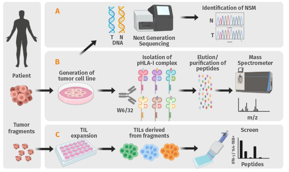
Mounting evidence shows that neoantigens play an important role in the clinical efficacy of cancer immunotherapies. Thanks to the support received from the BBVA Foundation's Comprehensive Program of Cancer Immunotherapy & Immunology – CAIMI-I and CAIMI-II, as well as other funding agencies, and through our group’s long-standing collaboration with Elena Garralda, Group Leader of VHIO’s Early Clinical Drug Development Group, and Director of our Research Unit for Molecular Therapy of Cancer (UTIM) – CaixaResearch, we are conducting a phase I clinical trial to evaluate the safety and tolerability of neoantigen-selected TILs.
This ongoing clinical trial is funded by the Instituto de Salud Carlos III – ISCIII (Carlos III Health Institute) and adopts a highly personalized approach (Figure) to screen for T-cell mediated recognition of mutated antigens. In this pilot study we aim to treat up to 10 patients with epithelial cancers and melanoma refractory to standard therapies. By enriching for neoantigen-reactive lymphocytes, we hope to extend the efficacy of TIL therapy beyond melanoma. In addition, our group is involved in several other clinical trials using non-selected ex vivo expanded TIL for the treatment of melanoma, germ cell cancers and tumors with mutations in the SWI/SNF complex.
One of our group’s strategic goals is to develop strategies that exploit the circulating tumor-reactive lymphocyte repertoire. We have shown that tumor-reactive T cells can frequently be detected circulating in the blood of cancer patients, irrespective of the specific tumor type. The ability to track and monitor tumor-reactive CD8+ and CD4+ T cells in blood has tremendous therapeutic potential, but this approach is more challenging due to the lower prevalence of these cells. We have contributed to defining a phenotypic signature that can guide the identification and enrichment of tumor and neoantigen-reactive T cells from the blood of cancer patients.
We have also demonstrated the use of circulating tumor DNA (ctDNA) to identify neoantigens, offering a minimally invasive approach to analyzing tumor-specific antigens. We are also developing a computational pipeline to conduct extensive profiling of the peripheral blood T-cell response at the single-cell level in patients treated with immune checkpoint inhibitors (ICIs) and vaccines to better understand the determinants of treatment response.
Our group is also interested in developing high-throughput technologies to better decipher the landscape of tumor antigens that increase cancer cells’ susceptibility to immune attack, and advance insights into shared neoantigens as well as mechanisms of resistance to T-cell cytotoxicity.
- Leverage peripheral blood to detect neoantigens and reactive T cells that can be used for therapeutic interventions.
- Track tumor antigen-specific T cell immune dynamics in patients treated with immunotherapy to understand determinants of response and toxicity.
- Establish a screening platform to identify novel sources of tumor antigens that contribute to tumor immunogenicity.
- Isolate and characterize T-cell receptors (TCRs) targeting public neoantigens and cancer germline antigens for the development of TCR gene-engineered therapies.
- Investigate tumor cell intrinsic mechanisms of resistance to T-cell mediated cytotoxicity.
- We identified 3 TCRs targeting public neoantigens derived from PIK3CA and TP53 using our proprietary neoepitope discovery platform.
- We contributed to shedding light on how chemotherapy induces the expression of PD-L2 in senescent cells, thereby facilitating immune evasion.
- Led by Elena Garralda, Head of VHIO’s Early Clinical Drug Development Group, we collaborate in the EU-Funded PragmaTIL multicenter clinical trial. Patients will be randomized to either TIL with high-dose interleukin 2 (HD-IL-2) or ANV419, an IL-2 receptor agonist.
- Conduct clinical scale validation of a first-generation T-cell product derived from peripheral blood of cancer patients.
- Chaib S, López-Domínguez JA, Lalinde-Gutiérrez M, Prats N, Marin I, Boix O, García-Garijo A, Meyer K, Muñoz MI, Aguilera M, Mateo L, Stephan-Otto Attolini C, Llanos S, Pérez-Ramos S, Escorihuela M, Al-Shahrour F, Cash TP, Tchkonia T, Kirkland JL, Abad M, Gros A, Arribas J, Serrano M. The efficacy of chemotherapy is limited by intratumoral senescent cells expressing PD-L2. Nat Cancer. 2024 Mar;5(3):448-462.
- Martin-Liberal J, Garralda E, García-Donas J, Soto-Castillo JJ, Mussetti A, Codony C, Martin-Lluesma S, Muñoz S, Galvao V, Lostes J, Rotxes M, Prat-Vidal C, Palomero J, Muñoz A, Moreno R, García Del Muro X, Sureda A, Alemany R, Gros A, Piulats JM. Clinical protocol phase II study of tumor infiltrating lymphocytes in advanced tumors with alterations in the SWI/SNF complex: the TILTS study. Future Oncol. 2024;20(32):2437-2445.
- Chaib S, López-Domínguez JA, Lalinde-Gutiérrez M, Prats N, Marin I, Boix O, García-Garijo A, Meyer K, Muñoz MI, Aguilera M, Mateo L, Stephan-Otto Attolini C, Llanos S, Pérez-Ramos S, Escorihuela M, Al-Shahrour F, Cash TP, Tchkonia T, Kirkland JL, Abad M, Gros A, Arribas J, Serrano M. The efficacy of chemotherapy is limited by intratumoral senescent cells expressing PD-L2. Nat Cancer. 2024 Mar;5(3):448-462. doi: 10.1038/s43018-023-00712-x. Epub 2024 Jan 24.
- Martin-Liberal J, Garralda E, García-Donas J, Soto-Castillo JJ, Mussetti A, Codony C, Martin-Lluesma S, Muñoz S, Galvao V, Lostes J, Rotxes M, Prat-Vidal C, Palomero J, Muñoz A, Moreno R, García Del Muro X, Sureda A, Alemany R, Gros A, Piulats JM. Clinical protocol phase II study of tumor infiltrating lymphocytes in advanced tumors with alterations in the SWI/SNF complex: the TILTS study. Future Oncol. 2024;20(32):2437-2445. doi: 10.1080/14796694.2024.2385287. Epub 2024 Aug 12.
- Grantor: Instituto de Salud Carlos III (ISCIII). Title: Terapia celular de próxima generación con TIL específicos de neoantígenos para pacientes con tumores resistentes a inhibidores de puntos de control inmunitario.
- Grantor: Fundación Fero (Beca Fero). Title: Non-invasive personalized T-cell therapies targeting recurrent hot spot driver mutations in cancer.
- Grantor: Fundación BBVA. Alena Gros is a member of the VHIO-BBVA Foundation Comprehensive Program of Cancer Immunotherapy & Immunology (CAIMI).
- Grantor: La Caixa Health Research. Title: Development of enabling technologies for T-cell immunotherapy of solid tumors.
- Grantor: Ministerio de Ciencia e Innovación. Title: Mining the molecular determinants of the personalized T-cell response in cancer patients to develop more effective immunotherapies.
- Grantor: Asociación Española Contra el Cáncer. Title: Harnessing TCR T cell Therapies to target recurrent mutations in PIK3CA and KRAS (PI-TCR ACT).
- Grantor: Ministerio de Ciencia, Innovación y Universidades. Title: Personalized T-cell therapies targeting neoantigens with enhanced efficacy and broad applicability (PersACT).
- Chaib S, López-Domínguez JA, Lalinde-Gutiérrez M, Prats N, Marin I, Boix O, García-Garijo A, Meyer K, Muñoz MI, Aguilera M, Mateo L, Stephan-Otto Attolini C, Llanos S, Pérez-Ramos S, Escorihuela M, Al-Shahrour F, Cash TP, Tchkonia T, Kirkland JL, Abad M, Gros A, Arribas J, Serrano M. The efficacy of chemotherapy is limited by intratumoral senescent cells expressing PD-L2. Nat Cancer. 2024 Mar;5(3):448-462. doi: 10.1038/s43018-023-00712-x. Epub 2024 Jan 24.
- Martin-Liberal J, Garralda E, García-Donas J, Soto-Castillo JJ, Mussetti A, Codony C, Martin-Lluesma S, Muñoz S, Galvao V, Lostes J, Rotxes M, Prat-Vidal C, Palomero J, Muñoz A, Moreno R, García Del Muro X, Sureda A, Alemany R, Gros A, Piulats JM. Clinical protocol phase II study of tumor infiltrating lymphocytes in advanced tumors with alterations in the SWI/SNF complex: the TILTS study. Future Oncol. 2024;20(32):2437-2445. doi: 10.1080/14796694.2024.2385287. Epub 2024 Aug 12.
- NCT05141474. Assessment of the Safety and Tolerability of ex Vivo Next-generation Neoantigen-selected Tumor-infiltrating Lymphocyte (TIL) Therapy in Advanced Epithelial Tumors and Immune Checkpoint Blockade (ICB) Resistant Solid Tumors (NEXTGENTIL-ACT).
- NCT06630611. Evaluation of a Pragmatic Approach to Adoptive Cell Therapy (ACT) Using an IL2 Analog (ANV419) vs High Dose IL2 After Tumor Infiltrating Lymphocytes (TIL) Therapy in Patients With Melanoma, NSCLC and Cervical Cancer (PragmaTIL) (PragmaTIL).

Group Leader Héctor G. Palmer Senior Investigator Isabel Puig Postdoctoral Fellows David Aguilar, Oriol Arqués, Belen Elguero, Jordi Martínez-Quintanilla, Lorena Ramirez PhD Students Peijin Jiang, Alex Mur, Candida Salvans Technicians Anna Alcántara, Laia Cabellos, Debora Cabot, Irene Chicote, Raquel Flores, Jordi Vergés Bioinformatican Gilles Flamen
Stem Cells and Cancer Group
We study the mechanisms that allow cancer cells to persist, evade anticancer therapies and drive disease progression. By integrating gene editing (CRISPR/Cas) with classical signaling biochemistry, we study these alterations in cancer cell lines, genetically modified mice, patient-derived organoids (PDOs), and xenografts (PDXs). Our goal is to determine their functional impact on treatment response and identify new therapeutic targets.
We participate in a multidisciplinary task force involving medical oncologists, surgeons, radiologists, and nurses to translate laboratory discoveries into clinical applications.
Main research lines include:
Tumor cell dormancy
We study the biology of cancer cell dormancy as a driver of chemoresistance, minimal residual disease, and relapse. We previously discovered a core epigenetic network that governs cancer cell dormancy (Puig et al. 2018), and are now investigating the function of TET2, DPPA3 (Cuesta et al. 2023), and other epigenetic and transcription factors in tumor cell dormancy.
Our group is advancing the development of drugs targeting drivers of cancer cell dormancy, including TET2 modulators, and defining novel biomarkers for detecting chemo-resistant dormant tumor cells.
To accelerate clinical translation, we founded ONIRIA Therapeutics, a VHIO spin-off company developing innovative dormancy-targeting anticancer therapies (https://www.oniriatherapeutics.com/).
Response to target-directed treatments
We collaborate with oncologists and pharmaceutical partners to identify molecular mechanisms underlying sensitivity or resistance to drugs blocking Wnt/beta-catenin, Notch, PI3K/AKT, EGFR/LGR5, BRAF/MEK/ERK, CDKs or hippo signaling pathways (Tenbaum et al. 2012; Puig et al. 2013; Capdevila et al. 2020; Lehal et al. 2020, Harmstron et al. 2021; Herpers et al. 2022).
Based on our discoveries, we are designing prescreening tests for selecting patients for enrolment in clinical trials. Importantly, our findings are guiding rational drug combinations to treat cancer patients with progressive disease in academic clinical trials.
Advanced preclinical cancer models
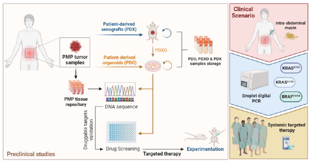
We are expanding and optimizing our PDX collection (colorectal cancer, neuroendocrine cancer, and peritoneal pseudomyxoma) to evaluate drug efficacy and metastatic potential using orthotopic injection and live imaging (positron emission tomography, computed tomography, and echography) approaches.
Our work is integrated into leading European consortia, including the EuroPDX, PERSIST-SEQ, CRCelerate, and the PMP Accelerate consortia, though which we contribute to advancing studies on cancer resistance and preclinical modeling.
- Study the role of epigenetic factors governing tumor cell dormancy in chemoresistance, minimal residual disease, relapse, dissemination and metastasis.
- Support ONIRIA Therapeutics spin-off company for the development of novel therapies that modulate cell dormancy and block cancer progression.
- Unveil the mechanisms of response to anticancer agents targeting the EGFR, BRAF, MEK, ERK, LGR5, Wnt, PARP or Hippo signaling pathways.
- Refine and expand our collection of patient-derived xenograft (PDX) and advanced cancer models and develop live imaging techniques.
- We have revealed key epigenetic factors governing cancer cell dormancy, hypoxia, chemoresistance and tumor recurrence, and developed effective small molecules targeting some of these processes.
- Our group has revealed new determinants of response to BRAF, MEK, Hippo, and Notch pathway inhibitors, and demonstrated the efficacy of new drug combinations targeting minimal residual disease in RET fusion-positive tumors.
- In collaboration with several European-funded networks, we have generated and refined advanced preclinical models of colorectal cancer.
We are committed to consolidating our spin-off company for developing drug modulators of cancer cell dormancy as a novel strategy to combat cancer.
We have already selected a lead compound with a low toxicity profile that activates TET2 and blocks tumor growth in advanced mouse models of cancer. Our first-in-class drug is ready to enter the preclinical regulatory phase. For this ambitious project, we have set up a team of experts in all stages of drug development.
Importantly, given VHIO’s successful track record in launching spin-off companies, ONIRIA is based at VHIO. Our researchers are evaluating first-in-class drugs in partnership with VHIO’s clinical investigators who have extensive expertise in early phase clinical studies.
Revealing new drivers of cancer cell dormancy
We aim to demonstrate the relevance of different epigenetic or transcription factors as master regulators of dormant tumor cells’ (DTC) distinctive phenotype. Specifically, our discovery of DPPA3 as a crucial factor in hypoxia and chemoresistance has opened many lines of investigation to explore its mechanisms of action and potential as a new drug target.
Supervised by our group’s senior investigators, our PhD students are currently studying other drivers governing cancer cell dormancy. This research is technically complex and relatively unexplored. Making a significant contribution to better understanding cancer cell dormancy and chemoresistance is particularly challenging. We aim to identify new drug targets and biomarkers to seek out and eradicate DTC and reduce chemoresistance and the risk of disease relapse.
Defining new effective therapies based on rational drug combinations
We have identified novel mechanisms of resistance to EGFR, BRAF, MEK, Wnt, Notch, RET, LGR5 or PARP inhibitors and will design and evaluate novel drug combinations that could benefit patients whose disease has progressed to first-line treatments. Based on the analyses of paired patients’ tumor samples and PDX models treated with matched therapies, this approach is already showing great promise.
We expect to report new insights into the mechanisms of action of some targeted drugs that will help guide oncologists to design and assess more precise and effective treatments. Within the framework of a recently granted EU IMI Innovative Medicines Initiative project, we will perform single cell multiomics analyses to evaluate drug resistance in our PDX models and fresh tissue samples from patients.
Innovating preclinical cancer models
One of our challenges is to integrate several imaging techniques (positron emission tomography, computed tomography (PET-CT) and echography) for the longitudinal evaluation of tumor growth in mice orthotopically implanted with patient-derived cells or genetically modified mouse models in response to new drug combinations. These comprehensive models could more faithfully recapitulate advanced disease in cancer patients, and therefore constitute a powerful tool in translating our laboratory results to the clinic.
- Martínez-Quintanilla J, Cabot D, Sabia D, Arqués O; PMPnet Group; Vergés J, Chicote I, Bijelic L; PMPnet Group; Cabellos L, Alcántara AM, Ramos I, Barrios P, Crusellas O, Palacio LM; PMPnet Group; Cámara JA, Barriuso J; PMPnet Group; Jiménez JJ, Muñoz-Torres P, Nonell L, Flores R, Médico E, Guaglio M; PMPnet Group; Ros J, Élez E, Tabernero J, Aziz O, Deraco M, Palmer HG; PMPnet Group; PMPnet Group. Precision Oncology and Systemic Targeted Therapy in Pseudomyxoma Peritonei. Clin Cancer Res. 2024 Sep 13;30(18):4082-4099.
- Title: Therapeutic efficacy of BRAF PROTACs CFT1946 and CFT-30723 in BRAFV600E CRC preclinical models.
Grant: C4 Therapeutics.
Duration: 2024-2025.
PI: Héctor G. Palmer. - Title: Identification of T cell receptors targeting hotspot mutations in common driver oncogenes from tumor specimens of Mismatch repair proficient colorectal carcinoma patients.
Grant: AstraZeneca
Duration: 2024-2027.
PI: Héctor G. Palmer. - Title: Therapeutic efficacy of extrachromosomal DNA (ecDNA) inhibitors in colorectal cancer tumors.
Grant: Boundless Bio.
Duration: 2024-2026.
PI: Héctor G. Palmer. - Title: Development and clinical validation of a CDX for ONR-001 monitoring in melanoma patients using plasmonic nanoparticles.
Grant: Ministerio de Ciencia, Innovación y Universidades.
Duration: 01/11/2024 – 01/11/2028.
PI, co-PI: Jesus de la Fuente and Héctor G. Palmer. - Project: ONIRIA Therapeutics.
Grant: Asociación Española Contra el Cáncer (AECC).
Funding program: AECC IMPACTO.
Duration: 01/03/2024 – 01/03/2027.
PI: Héctor G. Palmer. - Title: Investigating the single cell heterogeneity in CRC tumours for revealing the molecular circuits responsible for drug resistance as novel targets to fight disease persistence.
Grant: Instituto de Salud Carlos III (ISCIII).
Funding program: Fondo de Investigación en Salud (FIS).
Duration: 01/01/2024 – 31/12/2026.
PI: Héctor G. Palmer. - Title: Development of TET2 activating drugs for the treatment of melanoma.
Grant: Ministerio de Ciencia, Innovación y Universidades.
Funding Program: Proyectos de colaboración Público-Privada.
Duration: 01/11/2023 – 01/11/2027.
PI: Héctor G. Palmer. - Title: PERSIST-SEQ. Building a reproducible single-cell experimental workflow to capture tumour drug persistence.
Grant: European Commission-IMI2
Funding Program: H2020-JTI-IMI2-2020
Duration: 01/03/2021 – 01/03/2026.
PI: Héctor G. Palmer. - Title: Applying AI to scale up the Personal Discovery Process-A world-first, full-genome, personalised patient clinical trial for cancer.
Grant: European Innovation Council.
Funding program: EIC Accelerator.
Code according to the funding entity: 190103877.
Duration: 01/03/2023 – 28/02/2026 - Title: ONIRIA Therapeutics – BStarUP Health project.
Degree of contribution: Coordinator of total project, network or consortium
Funding program: BStarUP Health.
Duration: 01/04/2023 – 31/12/2025.
PI: Héctor G. Palmer. - Title: ONIRIA Therapeutics – Mind the Gap project.
Grant: Fundación Botín.
Funding program: Mind the Gap.
Duration: 01/04/2023 – 31/12/2025.
PI: Héctor G. Palmer. - Title: Multi-target approach including anti-angiogenic agent to overcome treatment resistance in BRAF mutant metastatic colorectal setting. BRAVE.
Grant: Instituto de Salud Carlos III (ISCIII).
Funding program: Academic Clinical Trials Call.
Code according to the funding entity: ICI21/00097.
Duration: 01/01/2022 – 31/12/2025
PI, co-PI: Héctor G. Palmer, Elena Élez. - Title: Design and Synthesis of novel TET2 inhibitors and PROTACs.
Grant: Ministerio de Ciencia e Innovación
Funding program: Ayudas para contrato Torres y Quevedo.
Code according to the funding entity: PTQ2021-012030
Duration: 11/11/2022 – 11/11/2025
PI, co-PI: Héctor G. Palmer, Elsa Martínez. - Title: Validating 5hmC as a companion diagnostic test for TET2 activator development.
Grant: Ministerio de Ciencia e Innovación.
Funding program: Ayudas para contrato Torres y Quevedo.
Code according to the funding entity: PTQ2021-012030.
Duration: 11/11/2022 – 11/11/2025.
PI, co-PI: Héctor G. Palmer, David Aguilar. - Title: Subvenció Joves Innovadors CAT- Investigo.
Grant: AGENCIA DE GESTIO D’AJUTS UNIVERSITARIS I DE RECERCA.
Funding program: Subvenció Joves Innovadors CAT- Investigo.
Code according to the funding entity: 200012TC4.
Duration: 01/05/2023 – 30/04/2025.
PI: Héctor G. Palmer. - Title: Subvenció Joves Innovadors CAT- Investigo.
Grant: AGENCIA DE GESTIO D’AJUTS UNIVERSITARIS I DE RECERCA.
Funding program: Subvenció Joves Innovadors CAT- Investigo.
Code according to the funding entity: 200017TC1.
Duration: 01/05/2023 – 30/04/2025.
PI: Héctor G. Palmer. - Title: Subvenció Joves Innovadors CAT- Investigo.
Grant: AGENCIA DE GESTIO D’AJUTS UNIVERSITARIS I DE RECERCA.
Funding program: Subvenció Joves Innovadors CAT- Investigo.
Code according to the funding entity: 200017TC3.
Duration: 01/05/2023 – 30/04/2025.
PI: Héctor G. Palmer. - Title: Subvenció Joves Innovadors CAT- Investigo.
Grant: AGENCIA DE GESTIO D’AJUTS UNIVERSITARIS I DE RECERCA.
Funding program: Subvenció Joves Innovadors CAT- Investigo.
Code according to the funding entity: 200017TC2.
Duration: 01/05/2023 – 30/04/2025.
PI: Héctor G. Palmer. - Title: Deciphering the molecular mechanisms of tumour cell resistance to cancer therapies.
Grant: Fundación de la Asociación Española Contra el Cáncer.
Funding program: Investigador AECC.
Duration: 01/01/2021 – 01/01/2025.
PI, co-PI: Puig Boreil, Héctor G. Palmer. - Title: PLAN DE NEGOCIO ONIRIA THERAPEUTICS: MODULACIÓN DE LA LATENCIA CELULAR PARA SUPERAR LA PERSISTENCIA DEL CÁNCER DE COLON.
Grant: Centro para el Desarrollo Tecnológico Industrial.
Funding program: Neotec.
Duration: 02/05/2023 – 31/12/2024. - Title: EpiGold-G: Desenvolupament d’un biosensor ultrasensible per a la detecció de modificacions epigenètiques en biòpsia líquida.
Grant: Generalitat de Catalunya.
Funding program: STARTUP CAPITAL.
Duration: 01/01/2023 – 31/12/2024.
PI: Héctor G. Palmer. - Title: Fármacos activadores de TET2 para el tratamiento del cáncer.
Grant: Instituto de Salud Carlos III (ISCIII).
Funding program: Proyectos de Investigación en salud, modalidad Desarrollo Tecnológico en Salud.
Code according to the funding entity: DTS21/00169.
Duration: 01/01/2022 – 31/12/2024.
PI: Héctor G. Palmer. - Title: Accelerator Award. Pseudomixoma peritonei: building a European multicentric cohort to accelerate new therapeutic perspectives.
Grant: Asociación Española Contra el Cáncer (AECC).
Code according to the funding entity: C69358/A29365.
Duration: 01/01/2020 – 31/12/2024
PI, co-PIs: Bipasha Chakrabarty; Jorge Barriuso; Omer Aziz; Gareth Evans; Amin Rostami; David Thorntom; Alexander Mirnezami; María Isabel Ramos Bernadó; Jil Barber; Giovanna Chiorino; Sarah Ennis; Norman Carr; Patrick Caswell; Paolo Verderio; Faheez Mohamed; Marco Vaira; Manuella Gariboldi; Sarah O´Dwyer; Marcello Deraco; Enzo Medico; Héctor G. Palmer. - Title: Tumor microenvironment-derived factors in localized colon cancer: clinical impact and therapeutic implications.
Grant: Fundación de la Asociación Española Contra el Cáncer (AECC).
Funding program: Grupos Coordinados AECC.
Code according to the funding entity: GCAEC20030CERV
Duration: 01/10/2020 – 30/09/2024.
PI, co-PI: Héctor G. Palmer, Andrés Cervantes. - Title: Evaluating the BRAF inhibitor Compound Ia in CRC.
Grant: ROCHE.
Start date: 03/06/2024
Duration: 2 years
PI: Héctor G. Palmer. - Title: Therapeutic efficacy of Claudin-1 Antibody in solid tumors.
Grant: ALENTIS.
Start date: 05/02/2024.
PI: Héctor G. Palmer. - Title: Therapeutic targeting the Hippo Pathway in colorectal cancer.
Grant: Ikena Oncology.
Duration: 01/09/2022-01/08/2024.
PI: Héctor G. Palmer. - Title: Evaluating the efficacy of CDK2/9 and PLK-1 inhibitors in CRC.
Grant: Cyclacel Pharmaceuticals.
Start date: 01/01/2022.
Duration: 2 years.
PI: Héctor G. Palmer. - Grant: “la Caixa” Foundation.
Funding Program: Doctoral INPhINIT-Retaining Fellowships at research centers of Excellence in Spain and Portugal.
Duration: 2020-2024.
Investigator: Alex Mur. - Title: Evaluating the efficacy of CDK2/9 and PLK-1 inhibitors in CRC.
Grant: Cyclacel.
Duration: 2022-2025.
PI: Héctor G. Palmer. - Title: Accelerator Award. ACRCelerate: Colorectal Cancer Stratified Medicine Network.
Grant: Cancer Research UK (CRUK), Asociación Española contra el Cancer (AECC) & Associazione Italina per la Ricerca sul Cancro (AIRC).
Duration: 2018-2024.
PI: Héctor G. Palmer. Consortium coordinator: Prof. Owen Sansom.
- Multi-target approach including anti-angiogenic agent to overcome treatment resistance in BRAF mutant metastatic colorectal setting. BRAVE.
Funding entity: Instituto de Salud Carlos III (ISCIII)
Funding program: Academic Clinical Trials Call.
Code according to the funding entity: ICI21/00097.
Duration: 01/01/2022 – 31/12/2025.
PI, co-PI: Héctor G. Palmer, Elena Élez.

Group Leader Mate Maus PhD Students Francesca Cogo, Marc Guasch, Ning Huang
Aging and Cancer Group
Founded in January 2023 by Mate Maus, VHIO’s Aging and Cancer Group is dedicated to translating aging research into oncology practice. We serve cancer patients, elderly individuals at risk of developing cancer, and cancer survivors facing accelerated health decline. We achieve this by developing innovative technologies and therapeutics capable of exposing and targeting age-related alterations in the metabolism of iron and other metals within the tissue microenvironment, recognized as drivers of malignancies.
To achieve our goals, we leverage state-of-the-art screening technologies, along with in vivo and in vitro models of cancer, anti-tumor immunity and aging. Collaborating with clinical oncologists, we strive to enhance our understanding of how the metabolic aging of the tissue microenvironment promotes cancer and hinders anti-tumor immunity. Our vision is a future where non-invasive metabolic imaging enables early detection of high-risk sites for cancer development, and therapeutic interventions that target the metabolic bottlenecks associated with aging that rejuvenate the tissue microenvironment and restore anti-tumor immunity to eliminate cancer.
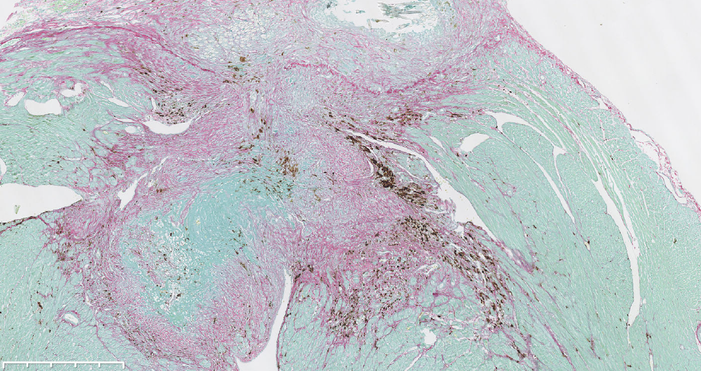
Tumorigenesis in the Metabolically Aged Tissue Microenvironment. The connection between aging and cancer extends beyond the accumulation of mutations. Mounting evidence suggests that the aging tissue microenvironment actively selects and promotes the progression of mutant cells toward malignancy. Building on our experience in exploring the interplay between inflammation and tumorigenesis (Saint-Fleur* and Maus* et al. Cell Rep. 2018), and our recent findings (Maus* and López-Polo* Nat Metab. 2023), we investigate how renewing tissues adapt to age-related disruption in iron metabolism and how this adaptation contributes to alterations in the differentiation trajectory and fitness landscape of precancerous cells in both bone marrow and the colon, pushing them toward malignancy. Our goal is to develop non-invasive methods capable of identifying high-risk sites for cancer development by imaging disruption in iron metabolism. Additionally, we aim to devise therapeutic strategies that can rejuvenate the tissue microenvironment by restoring iron metabolism, thereby offering a promising approach to treat cancer.
The Metabolism of Cancer Survivorship. As advances in oncology lead to a growing population of cancer survivors, the downsides of cancer therapies are becoming increasingly apparent. Cancer survivors not only face a shortened life expectancy but also grapple with higher rates of inflammatory and fibrotic diseases compared to the general population. Drawing on our experience in studying metabolism (Maus et al. Cell Metab. 2017) and our recent findings (Maus et al. Nat Metab. 2023), our focus is on unraveling the link between the adverse effects of cytotoxic therapies and iron accumulation. In this research line, our goal is to shed light on how cancer therapy disrupts both systemic and cellular iron homeostasis, and understand the intricate links to cellular senescence, fibrosis and accelerated aging in individuals who have received treatment with cytotoxic therapies. Ultimately, our goal is to develop innovative diagnostics and therapeutics that can enhance the quality of life for cancer survivors.
The Metabolic Aging of Anti-Tumor Immunity. The decline in anti-tumor immunity in aging is recognized as a key contributor to the elevated incidence of cancer in the elderly. Leveraging our expertise in studying immunometabolism (Vaeth* and Maus* et al. Immunity. 2017), and the metabolism of aging (Maus et al. Nat Metab. 2023), our research in this area focuses on investigating how age-related disruption in iron metabolism is associated with decreased tumor surveillance and T cell exhaustion. Our objective is to identify strategies that can rejuvenate the metabolic microenvironment, aiming to reinvigorate anti-tumor immunity.
*Equal contribution.
- Marc and Ning presented their research at the 1st International Conference of the Spanish Society of Cellular Senescence, SENESCEL, 07-08 October, Santiago de Compostela, Galicia (Spain).
- Britt Van Voskuijlen, our first Master’s Student, defended her thesis at Utrecht University in the Netherlands. Her research focused on targeting the metabolic profile of senescent cells to mitigate the profibrotic side effects of DNA-damaging cancer therapies.
- Our lab received the prestigious FERO Foundation Award for studying early-onset cancers, and the ”la Caixa” Foundation Innovation Award to develop drugs targeting the late effects of cancer treatment.
- We have expanded our research capabilities to investigate metabolic bottlenecks as drivers of age-related decline in antitumor immunity through establishing in silico and in vitro studies, as well as analyzing human tissue samples and performing in vivo experiments.
- Two impactful research papers published in Nature Communications and Cell Death and Disease.
- In 2024, we celebrated our lab's second anniversary.
- Expanding experimental data: Generate and analyze new in vivo and in vitro datasets to advance our research.
- Exploring tumor immunology: Deepen our understanding of tumor immunology in a metabolically aged environment and its implications for cancer progression and therapy.
- Advancing immunometabolism: Investigate the intersection of immune cell metabolism and aging to uncover novel therapeutic targets.
- Expansion of our team: Recruit additional team members, including postdocs, PhD students, and MSc students, to strengthen our lab’s expertise and capacity.
- Secure additional funding: Apply for new grants to support innovative projects and long-term goals.
- Consolidation of our lab: Establish a strong foundation for sustained research excellence and collaboration.
- Outreach and engagement: Increase our scientific impact through publications, presentations, and community engagement.
- Team building: Continue to foster a collaborative, creative, and supportive lab culture.
- López-Polo V, Maus M, Zacharioudakis E, Lafarga M, Attolini CS, Marques FDM, Kovatcheva M, Gavathiotis E, Serrano M. Release of mitochondrial dsRNA into the cytosol is a key driver of the inflammatory phenotype of senescent cells. Nat Commun. 2024 Aug 27;15(1):7378.
- XXVIII FERO Foundation Award
Project title: Colon Metabolism and Cancer Promotion: developing early detection strategies for young adults. Duration: 2025-2026.
PI: Mate Maus. - CaixaResearch Institute Innovation Hub
Project title: Treating age and cancer therapy-associated fibrotic diseases by targeting senescence associated iron accumulation. Duration: 2024-2025.
PI: Mate Maus.
- This group is supported by the ”la Caixa” Foundation - CaixaResearch.
- Ayudas para contratos Ramón y Cajal (RYC) (REF: RYC2020-030652-I).
- José Baselga Innovative Disruption Program (Astra Zeneca Foundation; REF: 10050754).
- Generacion de Conocimiento (Spanish Ministry of Science; REF: PID2022-142205OB-I00).
- La Caixa Hub Innovation Award.
- XXVIII Fundacion FERO; (FERO Foundation; REF: BFERO2024.03)
- Amengual J, Alay A, Vaquero J, Gonzalez-Sanchez E, Bertran E, Sánchez A, Herrera B, Meyer K, Maus M, Serrano M, Martínez-Chantar ML, Fabregat I. Iron chelation as a new therapeutic approach to prevent senescence and liver fibrosis progression. Cell Death Dis. 2024 Sep 17;15(9):680. doi: 10.1038/s41419-024-07063-0.
- López-Polo V, Maus M, Zacharioudakis E, Lafarga M, Attolini CS, Marques FDM, Kovatcheva M, Gavathiotis E, Serrano M. Release of mitochondrial dsRNA into the cytosol is a key driver of the inflammatory phenotype of senescent cells. Nat Commun. 2024 Aug 27;15(1):7378. doi: 10.1038/s41467-024-51363-0.

Group Leader Joan Seoane Staff Scientist Ignasi Barba Postdoctoral Fellows Ester Bonfill-Teixidor, Laia Cuesta, Clara Durán, Sergio Espinosa, Raffaella Iurlaro, Nicolás Pelaez Graduate Students Laura Carrillo, Cayetano Galera, María López, Almudena Neva Lab Manager Alexandra Arias Technicians Isabel Cuartas, María Duarte
Gene Expression and Cancer Group
We are committed to developing novel therapeutic compounds to improve cancer treatment. Our recent work characterized the preclinical properties and mechanism of action of a LIF-targeting agent in cervical cancer (Bonfill-Teixidor et al. 2024) (Figure). Our study showed that human papilloma virus (HPV) induces LIF expression via the NFKB pathway. In turn, LIF suppresses type I IFN and CXCL9 secretion, thereby preventing an effective anti-tumor immune response. These findings suggest that cervical cancer could be a promising indication for clinical trials combining anti-LIF compounds with immune checkpoint inhibitors.
MSC-1/AZD0171, a LIF-neutralizing antibody, was developed by VHIO spin-off Mosaic Biomedicals (co-founded by Joan in 2012). Mosaic Biomedicals was acquired by Medimmune/AstraZeneca in 2020. A phase II study evaluating this compound in metastatic pancreatic adenocarcinoma has been completed, with data maturation pending (NCT04999969). Additionally, a new clinical trial testing AZD0171 in non-small cell lung cancer opened in 2023 and was actively recruiting patients in 2024 (NCT05061550).
The role of B cell is not fully understood in the context of the anti-cancer immune response. We have characterized the B cell clonality in breast tumors by sequencing the BCR. We have found that the B cell clonality has an important impact on the immunosurveillance of breast cancer (Sammut et al. 2024).
Moreover, in collaboration with Roche, we have contributed to the discovery and preclinical characterization of a novel compound to complement the treatment of MSI-high colorectal cancer. This selective allosteric inhibitor of WRN helicase showed efficacy in preclinical models of MSI colorectal cancer (Baltgalvis et al. 2024), setting the rationale for advancing into evaluation in clinical trials.
Glioblastoma (GBM) and brain metastasis remain among the most aggressive of all cancers with a dismal prognosis and limited therapeutic options. Metabolism in GBM exhibits some particularities that could be considered a therapeutic target. We have summarized the latest research in this area in a recent review article (Barba et al. 2024).
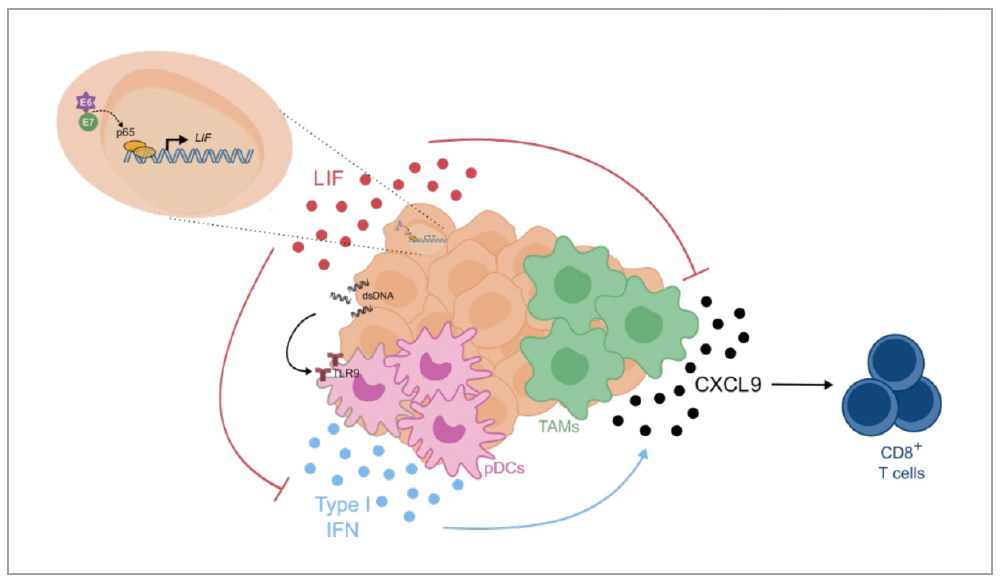
- Identify novel immunotherapeutic targets and biomarkers to improve therapy response prediction.
- Explore the tumor microenvironment to uncover mechanisms of immune regulation.
- Develop non-invasive molecular diagnostic methods for detecting circulating immune and non-immune cancer biomarkers.
- Generate innovative patient-derived models to advance immunotherapy research.
- We have revealed key epigenetic factors governing cancer cell dormancy, hypoxia, chemoresistance and tumor recurrence, and developed effective small molecules targeting some of these processes.
- Our group has revealed new determinants of response to BRAF, MEK, Hippo, and Notch pathway inhibitors, and demonstrated the efficacy of new drug combinations targeting minimal residual disease in RET fusion-positive tumors.
- In collaboration with several European-funded networks, we have generated and refined advanced preclinical models of colorectal cancer.
- Identify novel immunotherapeutic targets.
- Deepen understanding of the tumor microenvironment and immune system.
- Develop and implement non-invasive liquid biopsy methods to characterize the brain tumor immune landscape in clinical practice.
- Bonfill-Teixidor E, Neva-Alejo A, Arias A, Cuartas I, Iurlaro R, Planas-Rigol E, Solé L, Pecharromán I, Cabrera S, García Á, Garcia-Illescas D, Espinosa L, Gil-Moreno A, Oaknin A, Seoane J. Cervical Cancer Evades the Host Immune System through the Inhibition of Type I Interferon and CXCL9 by LIF. Clin Cancer Res. 2024 Oct 1;30(19):4505-4516.
- Sammut SJ, Galson JD, Minter R, Sun B, Chin SF, De Mattos-Arruda L, Finch DK, Schätzle S, Dias J, Rueda OM, Seoane J, Osbourn J, Caldas C, Bashford-Rogers RJM. Predictability of B cell clonal persistence and immunosurveillance in breast cancer. Nat Immunol. 2024 May;25(5):916-924.
- Baltgalvis KA, Lamb KN, Symons KT, Wu CC, Hoffman MA, Snead AN, Song X, Glaza T, Kikuchi S, Green JC, Rogness DC, Lam B, Rodriguez-Aguirre ME, Woody DR, Eissler CL, Rodiles S, Negron SM, Bernard SM, Tran E, Pollock J, Tabatabaei A, Contreras V, Williams HN, Pastuszka MK, Sigler JJ, Pettazzoni P, Rudolph MG, Classen M, Brugger D, Claiborne C, Plancher JM, Cuartas I, Seoane J, Burgess LE, Abraham RT, Weinstein DS, Simon GM, Patricelli MP, Kinsella TM. Chemoproteomic discovery of a covalent allosteric inhibitor of WRN helicase. Nature. 2024 May;629(8011):435-442.
- Title: Exploring the feasibility of predictive and pharmacodynamics biomarkers of immunotherapy in solid tumors (Immune-4ALL).
Funding: Institute of Health Carlos III-IMPACT.
PI: Joan Seoane.
Funding period: 2023-2026. - Title: GNOT (Grup de Neuro-Oncologia Translacional).
Funding: Agència de Gestió d’Ajuts Universitaris i de Recerca (AGAUR) i Servei General de Recerca (SGR).
PI: Joan Seoane.
Funding period: 2023-2026. - Title: Estudio del microambiente inmunosupresor del tumor cerebral.
Funding: Institute of Health Carlos III-FIS.
PI: Joan Seoane.
Funding period: 2023-2025. - Title: Biopsia Líquida para la caracterización y monitorización de la metástasis leptomeníngea.
Funding: Contigo Foundation.
PI: Joan Seoane.
Funding period: 2024. - Title: Evaluating the therapeutic potential of immunosuppressive paracrine cytokines in the tumor microenvironment of metastatic lesions.
Funding: European Comission ERA-NET TRANSCAN-3.
Type of participation: Coordinator.
Funding period: 2023-2025. - Title: Generate a novel of patient-derived ex vivo tumor platform that recapitulates the native tumor microenvironment of cancer patients.
Funding: JBID Project AstraZeneca.
PI: Joan Seoane.
Funding period: 2023-2026. - Title: Identification of biomarkers to predict response to LIF inhibitors and Search for therapeutic compounds to combine with anti-LIF agents.
Funding: AstraZeneca.
PI: Joan Seoane.
Funding Period: 2022-2024.
- Bonfill-Teixidor E, Neva-Alejo A, Arias A, Cuartas I, Iurlaro R, Planas-Rigol E, Solé L, Pecharromán I, Cabrera S, García Á, Garcia-Illescas D, Espinosa L, Gil-Moreno A, Oaknin A, Seoane J. Cervical Cancer Evades the Host Immune System through the Inhibition of Type I Interferon and CXCL9 by LIF. Clin Cancer Res. 2024 Oct 1;30(19):4505-4516. doi: 10.1158/1078-0432.CCR-24-0385.
- Sammut SJ, Galson JD, Minter R, Sun B, Chin SF, De Mattos-Arruda L, Finch DK, Schätzle S, Dias J, Rueda OM, Seoane J, Osbourn J, Caldas C, Bashford-Rogers RJM. Predictability of B cell clonal persistence and immunosurveillance in breast cancer. Nat Immunol. 2024 May;25(5):916-924. doi: 10.1038/s41590-024-01821-0.
- Baltgalvis KA, Lamb KN, Symons KT, Wu CC, Hoffman MA, Snead AN, Song X, Glaza T, Kikuchi S, Green JC, Rogness DC, Lam B, Rodriguez-Aguirre ME, Woody DR, Eissler CL, Rodiles S, Negron SM, Bernard SM, Tran E, Pollock J, Tabatabaei A, Contreras V, Williams HN, Pastuszka MK, Sigler JJ, Pettazzoni P, Rudolph MG, Classen M, Brugger D, Claiborne C, Plancher JM, Cuartas I, Seoane J, Burgess LE, Abraham RT, Weinstein DS, Simon GM, Patricelli MP, Kinsella TM. Chemoproteomic discovery of a covalent allosteric inhibitor of WRN helicase. Nature. 2024 May;629(8011):435-442. doi: 10.1038/s41586-024-07318-y.
- Barba I, Carrillo-Bosch L, Seoane J. Targeting the Warburg Effect in Cancer: Where Do We Stand? Int J Mol Sci. 2024 Mar 8;25(6):3142. doi: 10.3390/ijms25063142.
- Involved in NCT05187624 EGFRvIII TCB phase I.
- Involved in ISRCTN13713551 BRAF inhibitor phase I.
- Involved in NCT04999969, NCT05061550, MSC1/AZD0171 phase II trials.

Group Leader Francisco Martínez Jiménez Data Analyst Joseph Usset Postdoctoral Fellows Ana Dueso Barroso, Manuel Jara Espejo, Lucas Michel Todó PhD Students Marc Arbones (co-supervised), Yulia Kremlyakova (co-supervised), Natasa Mortvanski Junior Bioinformatician Sergio Esteban Echeverría Master’s Student Jone Gutierrez
Computational Immunogenomics Group
Our group focuses on advancing insights into the genomic basis of tumorigenesis and its interplay with the immune system. We perform large-scale tumor genomic analyses matched with a comprehensive profiling of the tumor immune microenvironment to better understand the tumor-immune crosstalk. Our final goal is to leverage this knowledge to improve the clinical development of tailored immunotherapies.
Cancer is characterized by an uncontrolled growth of cells that are not cleared by the immune system. Every tumor has a unique composition of genomic alterations acquired over the evolution of the tumorigenic clone. While tumors often harbor hundreds to thousands of genomic alterations, only a handful are responsible for the tumorigenic process (so-called drivers). Driver alterations tend to be cancer-type specific (Martínez-Jiménez et al. 2020) and are generally conserved between primary and metastatic lesions (Martínez-Jiménez et al. 2023).
It is however becoming increasingly clear that tumor genomic alterations are not the only promoters of tumor development and metastasis. Other factors including cancer immune escape also play a critical role. In this context, we are interested in developing computational models to 1) further understand the interplay between tumor evolution and the immune system, and 2) harness this knowledge to improve the clinical development of tailored immunotherapies.
Our approach is based on integrating the genomic portrait of tumors (based on whole-exome/genome sequencing), which includes the identification of simple and complex tumor specific alterations, rationalized prioritization of tumor neoantigens, and characterization of tumor HLA-I status and other immune escape alterations; combined with a quantitative portrayal of the tumor immune microenvironment (using bulk transcriptomics/proteomics immune deconvolution, single cell and geospatial immune profiling, and TCR-sequencing). As an example, we have recently performed a large-scale analysis of the prevalence and impact of genetic immune escape alterations across >6,000 primary and metastatic tumors (Martinez-Jiménez, Priestley et al. 2023).
Thanks to our close collaboration with the Hartwig Medical Foundation we have access to one of the largest resources of whole genome sequenced tumors and full tumor transcriptomes (currently >7,000 patients, https://database.hartwigmedicalfoundation.nl/). Moreover, in-house data is generated to support the validation of our analyses and address specific questions that require more targeted analysis.
Since our VHIO group was established in February 2023, we have obtained competitive funding to support multiple projects, participated in several national and international conferences, and we published five peer-reviewed papers in top journals.
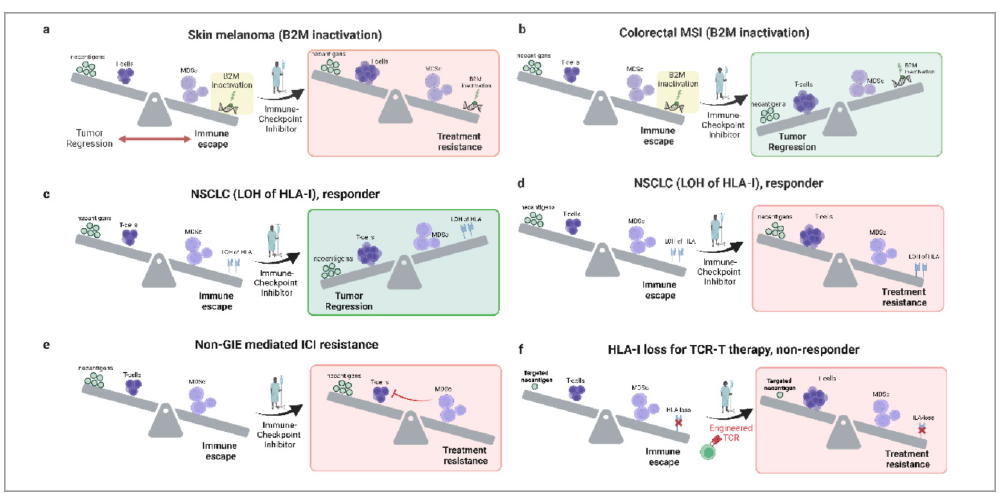
- Study the immune-oncogenicity trade-off of cancer mutations.
- Understand the distinctive genomic and transcriptomic traits of cancer of unknown primary (CUP).
- Understand the transcriptional reprograming of metastatic tumors and the interplay with the genomic background
- Co-lead cancer genomics projects in collaboration with the Hartwig Medical Foundation:
- Identify new clinical biomarkers for non-responders to cancer therapies.
- Demonstrate the clinical added value of WGS as part of routine care.
- Four new researchers joined our group in 2024.
- We have consolidated our collaboration with the Hartwig Medical Foundation.
- Published a Trends in Cancer article as corresponding author about the origin and timing of genetic immune escape events in cancer.
- We have obtained three competitive research grants to support our research.
- Our investigators have participated at multiple national and international meetings including the 19th Spanish Association for Cancer Research (ASEICA) International Congress and the European Society for Medical Oncology’s (ESMO) Molecular Analysis for Precision Oncology Congress (MAP 2024).
- Understand the transcriptional reprograming of metastatic tumors and the interplay with the tumor genomic background
- Decipher the trade-off between oncogenicity and immunogenicity of cancer mutations.
- Understand the role of HLA-II cancer cell expression in tumor evolution and metastasis
- Unravel the mysterious immunogenomic determinants of cancer of unknown primary (CUP).
- Expand our research group by securing international competitive funding.
- Engage in new synergistic projects with clinical teams at VHIO.
- Further develop our research collaboration with the Hartwig Medical Foundation.
- Martínez-Jiménez F, Chowell D. Genetic immune escape in cancer: timing and implications for treatment. Trends Cancer. 2024. doi: 10.1016/j.trecan.2024.11.002.
- Krishna C, Tervi A, Saffern M, Wilson EA, Yoo SK, Mars N, Roudko V, Cho BA, Jones SE, Vaninov N, Selvan ME, Gümüş ZH; FinnGen§; Lenz TL, Merad M, Boffetta P, Martínez-Jiménez F, Ollila HM, Samstein RM, Chowell D. An immunogenetic basis for lung cancer risk. Science. 2024 Feb 23;383(6685):eadi3808.
- Francisco Martínez Jiménez was awarded the 2024 CRIS Emerging Leader Award for the project entitled: Unravelling liver metastasis transcriptional regulators and their association to the tumor genomic landscape and the tissue of origin.
- Francisco Martínez Jiménez received a 2024 LAB AECC - Asociación Española Contra el Cáncer Award for the project: “Systematic characterization of distinctive immunogenomic traits of Cancers of Unknown Primary (CUPs) to identify novel therapeutic opportunities.
- Unravelling liver metastasis transcriptional regulators and their association to the tumor genomic landscape and the tissue of origin. CRIS Emerging Leader 2024. 2025-2029. Role: Principal Investigator
- Systematic characterization of distinctive immunogenomic traits of Cancers of Unknown Primary (CUPs) to identify novel therapeutic opportunities. Lab AECC 2024. November 2024-2027. Role: Principal
- Small cell lung cancer: from biology networks to tailored therapy. AECC 70 Survivorship 2024 Challenge. July 2024-2029. Group Funding: €250,000. Role: Work Package Leader: Comprehensive molecular and computational analysis”
- Ramon y Cajal program. Ministerio de Ciencia e Innovacion. (January 2024–2028). Principal Investigator: Francisco Martínez Jiménez (5th in Biomedicine). Characterization of the impact of genetic immune escape alterations on responses to Immune Checkpoint Inhibitors across human cancers
- Martínez-Jiménez F*, Chowell D*. Genetic immune escape in cancer: timing and implications for treatment. Trends in Cancer. doi: 10.1016/j.trecan.2024.11.002 Genetic immune escape in cancer: timing and implications for treatment. Trends Cancer. 2024 Dec 3:S2405-8033(24)00254-1. doi: 10.1016/j.trecan.2024.11.002. Epub ahead of print.
- Usset J, Rosendahl Huber A, Andrianova MA, Batlle E, Carles J, Cuppen E, Elez E, Felip E, Gómez-Rey M, Lo Giacco D, Martinez-Jimenez F, Muñoz-Couselo E, Siu LL, Tabernero J, Vivancos A, Muiños F, Gonzalez-Perez A, Lopez-Bigas N. Five latent factors underlie response to immunotherapy. Nat Genet. 2024 Oct;56(10):2112-2120. doi: 10.1038/s41588-024-01899-0. Epub 2024 Sep 12.
- Marcos-Villar L, Perdiguero B, Anthiya S, Borrajo ML, Lou G, Franceschini L, Esteban I, Sánchez-Cordón PJ, Zamora C, Sorzano CÓS, Jordá L, Codó L, Gelpí JL, Sisteré-Oró M, Meyerhans A, Thielemans K, Martínez-Jiménez F, López-Bigas N, García F, Alonso MJ, Plana M, Esteban M, Gómez CE. Modulating the immune response to SARS-CoV-2 by different nanocarriers delivering an mRNA expressing trimeric RBD of the spike protein: COVARNA Consortium. NPJ Vaccines. 2024 Mar 6;9(1):53. doi: 10.1038/s41541-024-00838-8.
- Krishna, C., Tervi, A., Saffern, M., Wilson, E. A., Yoo, S. K., Mars, N., Roudko, V., Cho, B. A., Jones, S. E., Vaninov, N., Selvan, M. E., Gümüş, Z. H., FinnGen§, Lenz, T. L., Merad, M., Boffetta, P., Martínez-Jiménez, F., Ollila, H. M., Samstein, R. M., & Chowell, D. (2024). An immunogenetic basis for lung cancer risk. Science. 2024 Feb 23;383(6685):eadi3808. doi: https://doi.org/10.1126/science.adi3808

Group Leader Francisco M. Barriga Lab Manager Xieng Chen Wang Postdoctoral Fellows Etna Abad, Dario Ruiz PhD Students Sofia Calpe, Golsa Jahangiri, Matea Katić, Claudia Yáñez
Cancer Genome Engineering Group
Our group studies the function of large-scale chromosomal changes known as copy number alterations (CNAs). We combine state-of-the-art genome engineering strategies to uncover the mechanisms by which CNAs enable cancer cells to propagate and resist therapies. We are particularly interested in the role of these alterations in immune surveillance, tumor heterogeneity, and cancer genome evolution. Our long-term goal is to understand the complex biology of CNAs to identify new therapeutic strategies that target cells with these chromosomal aberrations.
Cancers arise through genetic and epigenetic alterations that drive the transformation of single cells into malignant tumors. Among the most frequent genetic changes observed in cancer are CNAs. As their name implies, CNAs change how many copies of specific regions of DNA a cancer cell has. Importantly, some CNAs are associated with poor clinical outcomes, yet we still do not know how they change the properties of tumor cells. To better understand the function of these alterations, our lab combines in vivo models of cancer with MACHETE, a new technique for engineering CNAs.
Our previous work focused on studying the co-deletion of a cluster of 17 type I interferons linked to the tumor suppressor genes CDKN2A/B. Using MACHETE (Molecular Alteration of Chromosomes with Engineered Tandem Elements), we showed that combined loss of the interferon cluster and tumor suppressors led to immune evasion, metastasis, and reduced response to immunotherapy compared to CDKN2A/B-only deletions (Barriga, Tsanov et al. 2022). This study illustrates the complex biology of CNAs and underscores the need for precise modeling approaches to elucidate the biological consequences of these alterations. We also recently published a protocol paper (Barriga and Lowe, 2024), to facilitate the establishment of MACHETE in laboratories interested in creating megabase-sized genomic deletions. From a broader perspective, CNAs have a unique operational logic rooted in changes of gene dosage and DNA topology, which we aim to explore in greater depth.
Beyond our work on CNAs, we are also developing new combinatorial genetic tools to dissect the complex cellular states in vitro and in vivo.
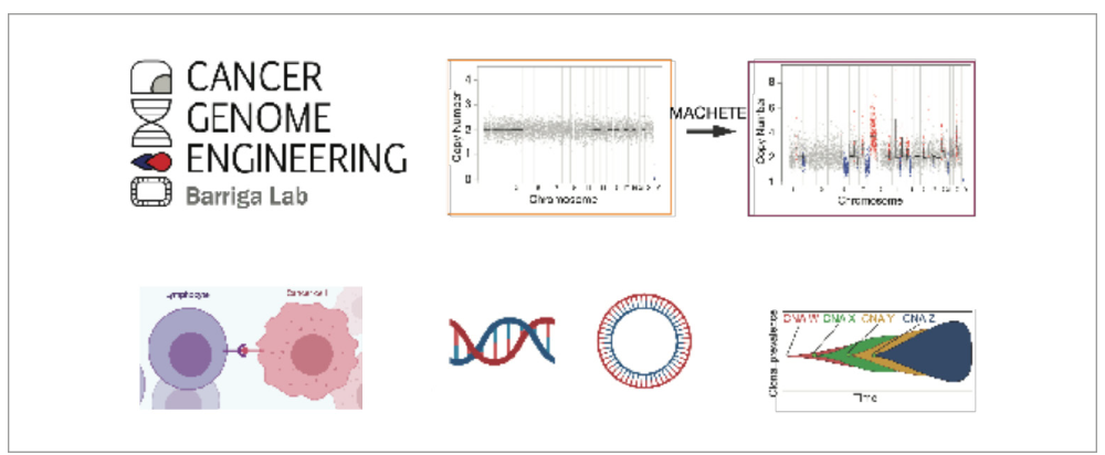
- Understand the role of CNAs as mechanisms of immune evasion and epigenetic rewiring.
- Identify the effect of CNAs in tumor heterogeneity.
- Dissect the contribution of CNAs and ploidy in shaping cancer genome evolution.
- Decipher the biology of unique cell states via combinatorial genetics models in vivo.
- Francisco Barriga was awarded a Ramón y Cajal Fellowship (Agencia Estatal de Investigación), and also received the XXVI FERO Fellowship.
- Dario Ruiz was awarded an Asociación Española Contra el Cáncer (AECC) Talent Postdoctoral Fellowship.
- A protocol article for genome engineering was published in Nature Protocols.
- Develop novel models to study genetic mechanisms of immune escape.
- Dissecting the genetic mechanisms of intra-tumor heterogeneity and genome evolution.
- The use of combinatorial genetics to dissect the biology of in vivo cell states.
- Barriga FM, Lowe SW. Engineering megabase-sized genomic deletions with MACHETE (Molecular Alteration of Chromosomes with Engineered Tandem Elements). Nat Protoc. 2024 May;19(5):1381-1399.
- Reference: 101041659 (Acronym: MACHETE)
Title: Unravelling the Functional Complexity of Cancer Genomes through Chromosome Engineering
Funding Entity – Call: ERC – Starting Grant
PI/Institution: Francisco Barriga, PhD / VHIO
Funding Period: January 2023 – December 2028 - Reference: RYC2022-037389-I
Title: Dissecting the function of copy number alterations in cancer.
Funding Entity – Call: Agencia Estatal de Investigación – Ayuda Ramón y Cajal 2022
PI/Institution: Francisco Barriga, PhD / VHIO
Funding Period: January 2024 – December 2028 - Reference: PID2022-141256OA-I00 (Acronym: EPI-MHC)
Title: Dissecting the epigenetic effects of MHC cluster deletion in pancreas cancer
Funding Entity – Call: Agencia Estatal de Investigación – Proyectos de Generación de Conocimiento 2022
PI/Institution: Francisco M. Barriga, PhD / VHIO
Funding Period: September 2023 – August 2026 - Reference: CNS2023-144041
Title: Unravelling the function of anti-tumor immune cell states through combinatorial genetics
Funding Entity – Call: Agencia Estatal de Investigación – Consolidación Investigadora 2023
PI/Institution: Francisco M. Barriga, PhD / VHIO
Funding Period: April 2024 – March 2026 - Reference: Beca FERO
Title: Dissecting the epigenetic and immune effects of MHC LOH in ovarian cancer
Funding Entity – Call: Fundación FERO – XXVI Beca FERO
PI/Institution: Francisco M. Barriga, PhD / VHIO
Funding Period: October 2024 – September 2026
- Barriga FM*, Lowe SW. Barriga FM, Lowe SW. Engineering megabase-sized genomic deletions with MACHETE (Molecular Alteration of Chromosomes with Engineered Tandem Elements). Nat Protoc. 2024 May;19(5):1381-1399. *Corresponding author.
- Tsanov KM, Barriga FM, Ho YJ, Alonso-Curbelo D, Livshits G, Koche RP, Baslan T, Simon J, Tian S, Wuest AN, Luan W, Wilkinson JE, Masilionis I, Dimitrova N, Iacobuzio-Donahue CA, Chaligné R, Pe'er D, Massagué J, Lowe SW. Metastatic site influences driver gene function in pancreatic cancer. bioRxiv [Preprint]. 2024 Mar 18:2024.03.17.585402.
- Zhao X, Liu B, William WN, Tsanov KM, Ho YJ, Barriga FM, Lim RJ, Trifas M, Du Y, Lowe SW, Dubinett SM, Davoli T, Lippman SM. Interferon-ε loss is elusive 9p21 link to immune-cold tumors, resistant to immune-checkpoint therapy and endogenous CXCL9/10 induction. J Thorac Oncol. 2024 Dec 24:S1556-0864(24)02539-5.

Group Leader José A. Seoane Postdoctoral Fellows Hubert Bretonniere, Lea Lemler, Silvana Maas Graduate Students Enric Álvarez, María Butjosa, María José Fariña, Arnau Llinàs, Laia Ollé, Juan Rafael Valera
Cancer Computational Biology Group
Headed by José A. Seoane, VHIO’s Cancer Computational Biology Group leverages epi(genetic) cancer datasets to identify the molecular mechanisms implicated in cancer initiation, progression, drug resistance and metastasis toward improving outcomes for patients.
We aim to advance insights into the role of chromatin regulatory elements in treatment response and metastasis, develop novel epigenetic synthetic lethality-based therapeutic options, improve patient stratification guided by multi-omics analysis, and discover novel epigenetic biomarkers of treatment response.
We have previously shown how the genetic modifiers of chromatin structure are associated with chemotherapy resistance in breast cancer (Seoane et al. 2019). Our group focuses on how these epigenetic alterations affect drug resistance and how epigenetic therapies can be used to target tumor suppressor genes.
We are also exploring how the machine learning-based horizontal and vertical integration of multi-omics datasets can facilitate the discovery of new cancer subgroups and biomarkers, as well as help to better predict treatment outcomes and drug response.
Our group has participated/participates in multiple international consortia including The Cancer Genome Atlas, the Human Tumor Atlas Network, Cancer Target Discovery and Development (CTD 2) Network, and, most recently, AURORA (metastatic breast cancer multi-omics cohort).
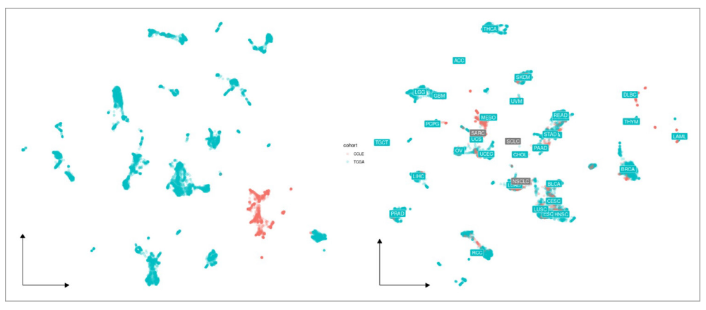
- Understand the role of chromatin regulatory elements in treatment response and metastasis.
- Discover new epigenetic biomarkers of drug response.
- Potentiate therapeutic options by combining epigenetic therapies with other agents.
- Discover potential environmental causes of early onset cancer.
- Develop clinically actionable machine learning algorithms for prediction and patient stratification.
- Our group is a member of the Vall d'Hebron TACTICS project. This four-year project was awarded with a FORTALECE grant from the Institute of Health Carlos III (ISCIII).
- In 2024, Huber Bretonniere and Juan Rafael Valera joined our team.
- Determine epigenetic synthetic lethality changes in gastrointestinal cancers.
- Investigate the epigenetic changes in post hormone treatment for ER+ and its association with metastatic breast cancer.
- Identify a cell-cycle based signature of treatment prediction in colon cancer.
- Identify exposome-related causes of cancer initiation.
- Develop clinically useful machine learning models
- Llinas-Bertran A, Bellet-Ezquerra M, Seoane JA. Epigenetic Control of Cancer Cell Dormancy and Awakening in Endocrine Therapy Resistance. Cancer Discov. 2024 May 1;14(5):704-706.
- Targeting synthetic lethality in Chromatin Regulatory Genes for gastrointestinal cancer treatment. Ref: PID2020-115097RA-I00. Agencia Estatal de Investigación. 9/2021-8/2025. PI: Jose A. Seoane.
- Ramon y Cajal Grant. Agencia Estatal de Investigación. 2021-2025.
- Epigenetic differences associated with hormone treatment resistant breast cancer heterogeneity. Fundación FERO. 2022-2024.
- Identificación in Silico de Respuesta a fármacos especifica de metastasis Entidad Financiadora: MCIN/AEI/10.13039/501100011033. Ref: CNS2022-135424. 01/09/2023 – 31/08/2025. PI: Jose Antonio Seoane
- TACTIC: Explorando soluciones a los retos de salud mediante ciencia disruptiva, terapias avanzadas y medicina de sistemas. Programa FORTALECE ISCIII REF: FORT23/00034. 2024-2027.
- Llinas-Bertran A, Bellet-Ezquerra M, Seoane JA. Epigenetic Control of Cancer Cell Dormancy and Awakening in Endocrine Therapy Resistance. Cancer Discov. 2024 May 1;14(5):704-706.
- Miera-Maluenda M, Pérez-Torres M, Mañas A, Rubio-San-Simón A, Butjosa-Espín M, Ruiz-Duran P, Seoane JA, Moreno L, Segura MF. Advances in the approaches used to repurpose drugs for neuroblastoma. Expert Opin Drug Discov. 2024 Nov;19(11):1309-1319.

Group Leader Raquel Perez-Lopez Postdoctoral Fellow Francesco Grussu PhD Students Alonso García, Athanasios Grigoriou, Olivia Prior, Anna Voronova, Daniel Navarro, Maria Balaguer, Carlos Macarro Students Bernat Argelich Laboratory Technician Marta Buetas Computer Scientists Adrià Marcos, Camilo Monreal Research Fellow Luz María Atlagich, Nikos Satikoglou Data Curator Christina Zatse
Radiomics Group
The VHIO Radiomics Group is dedicated to advancing precision oncology through the development and clinical translation of artificial intelligence (AI) and data integration tools, with a particular focus on medical imaging. Our vision is to harness the full potential of radiomics and computational modelling to transform cancer care, making diagnostics more accurate, treatments more personalized, and patient outcomes improved.
By combining expertise in engineering and machine learning, we work toward the discovery, validation, and clinical qualification of imaging biomarkers that inform and guide treatment decision-making. We are also deeply committed to supporting drug development by leveraging functional imaging techniques within clinical trials, enabling real-time insights into treatment response.
Through our interdisciplinary approach, strong collaborations, and commitment to innovation, the VHIO Radiomics Group aims to be at the forefront of computational oncology, shaping the future of cancer diagnostics and therapeutics.
In 2024, we were thrilled to welcome Marta Buetas as a new technician to our team. Marta has a solid background in biomedical engineering and valuable experience in medical image analysis, specifically in oncology. We are confident that her fresh perspective will enrich the group's work, and we eagerly anticipate her future doctoral studies within our team. Her potential for innovative ideas, proactive approach, and leadership in project management are qualities we believe will significantly benefit our research activities.
We are proud to announce that Alonso Garcia successfully defended his doctoral thesis at the end of 2024, earning the distinguished grade of magna cum laude. His outstanding research over the past four years has yielded excellent results, with publications in prestigious journals such as European Radiology and Cell Reports Medicine. Additionally, Alonso has been involved in multiple collaborations with both internal groups at VHIO and various national and international partners, further amplifying the impact of his work.
We also had the pleasure of hosting Bernat Alberich, a master's thesis student from the University of Girona. Bernat conducted exceptional research on applying deep learning models for the automatic detection and segmentation of bone metastases. His contributions showcased the promising potential of advanced computational techniques in oncological imaging.
This year, our team secured several important grants, both as Principal Investigators and collaborators. Among these, we successfully obtained funding from the Fondo de Investigaciones Sanitarias (FIS), enabling us to further advance our research initiatives.
Raquel Perez-Lopez received notable appointments this year, becoming the Head of the Cancer Core Europe (CCE) Imaging Task Force, member of the European Association for the Study of Liver disease (EASL) Artificial Intelligence Task Force and the Head of the Oncological Unit at Sociedad Española de Radiología Médica (SERAM). These prestigious roles underscore her leadership in the field of oncological imaging and her continued commitment to advancing the integration of artificial intelligence in cancer research.
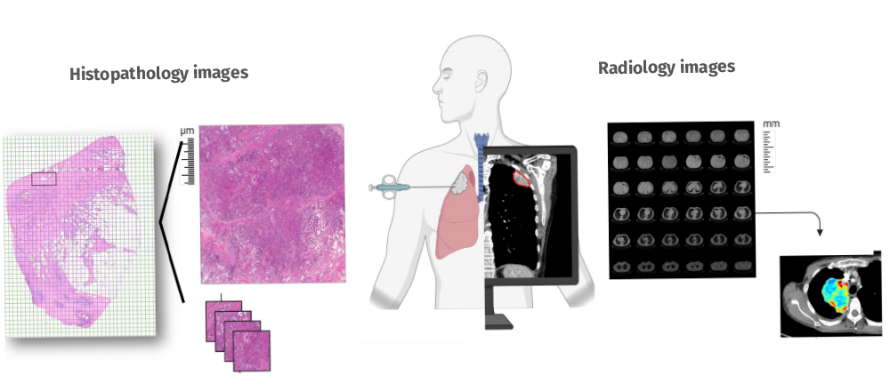
- Develop and optimize pipelines for AI models of data integration with particular focus on medical imaging and the integration of explainable models.
- Provide expertise in engineering and bioinformatics for the development and clinical qualification of imaging biomarkers for precision oncology to improve outcomes for cancer patients.
- Use functional imaging to optimize drug development in clinical trials.
- Integrate radiomics with other omics in translational studies to achieve a deeper understanding of tumor evolution and mechanisms of resistance to anti-cancer therapies.
- Our team published several high-impact articles in renowned journals such as Nature Reviews Cancer, Radiology: Artificial Intelligence, and Cell Reports Medicine. These publications advanced the field of radiomics and AI-driven imaging biomarkers in cancer.
- Raquel Perez-Lopez was appointed Head of the CCE Imaging Task Force, member of the EASL Artificial Intelligence Task Force and the Head of the Oncological Unit at SERAM, reflecting the team’s growing influence in oncological imaging.
- We have continued to expand existing partnerships with other groups and initiated new collaborative projects to promote the integration of imaging biomarkers in translational research projects.
- Continue to build a team incorporating talented radiologists, physicists, and engineers to develop research with cutting-edge imaging methods.
- Further promote the integration of imaging biomarkers in translational research projects at VHIO and other leading research centers.
- Develop novel imaging processing algorithms for advanced imaging.
- Perez-Lopez R, Ghaffari Laleh N, Mahmood F, Kather JN. A guide to artificial intelligence for cancer researchers. Nat Rev Cancer. 2024 Jun;24(6):427-441.
- Garcia-Ruiz A, Macarro C, Zacchi F, Morales-Barrera R, Grussu F, Casanova-Salas I, Sanguedolce F, Gonzalez M, Cresta-Morgado P, de Albert M, Garcia-Bennett J, Marmolejo D, Planas J, Roche S, Mast R, Zatse C, Piulats JM, Herrera-Imbroda B, Regis L, Agundez L, Olmos D, Calvo N, Escobar M, Carles J, Mateo J, Perez-Lopez R. Whole-body Magnetic Resonance Imaging as a Treatment Response Biomarker in Castration-resistant Prostate Cancer with Bone Metastases: The iPROMET Clinical Trial. Eur Urol. 2024 Sep;86(3):272-274.
- Garcia-Ruiz A, Pons-Escoda A, Grussu F, Naval-Baudin P, Monreal-Aguero C, Hermann G, Karunamuni R, Ligero M, Lopez-Rueda A, Oleaga L, Berbís MÁ, Cabrera-Zubizarreta A, Martin-Noguerol T, Luna A, Seibert TM, Majos C, Perez-Lopez R. An accessible deep learning tool for voxel-wise classification of brain malignancies from perfusion MRI. Cell Rep Med. 2024 Mar 19;5(3):101464.
- Prior O, Macarro C, Navarro V, Monreal C, Ligero M, Garcia-Ruiz A, Serna G, Simonetti S, Braña I, Vieito M, Escobar M, Capdevila J, Byrne AT, Dienstmann R, Toledo R, Nuciforo P, Garralda E, Grussu F, Bernatowicz K, Perez-Lopez R. Identification of Precise 3D CT Radiomics for Habitat Computation by Machine Learning in Cancer. Radiol Artif Intell. 2024 Mar;6(2):e230118. doi: 10.1148/ryai.230118. Erratum in: Radiol Artif Intell. 2024 May;6(3):e249001.
- Secured funding as Principal Investigators from the Fondo de Investigaciones Sanitarias (FIS) to support groundbreaking research in radiomics and imaging biomarkers.
- Established a significant collaboration with INCLIVA thanks to funding received from the Mutua Madrileña to support the GLADIATOR 2 study. This research focuses on advancing colon cancer treatment using artificial intelligence (AI)-powered data integration.
- We received the Spanish Society of Medical Radiology (SERAM) Award for Research in Radiology in recognition of our innovative contributions to the field of radiological imaging and artificial intelligence in cancer research.
We participate in several multicentric international projects which provide a unique opportunity to accelerate the development and application of novel AI-tools in cancer research, ultimately leading to advancements in clinical practice.
- Project title: ODELIA: An Open Consortium for Decentralized Medical Artificial Intelligence.
Co-Investigator: Raquel Perez.
Duration: 2023-2027.
The project aims to overcome the obstacles of data collection in healthcare by utilizing swarm learning, where partners work together to train artificial intelligence (AI) models without the need to share any personal patient data.
ODELIA seeks to overcome current boundaries in data sharing and accelerate the scale-up of medical AI in Europe for the benefit of European citizens, patients, and clinicians. Our group is collaborating in the development of a swarm learning model to enhance MRI screening in breast cancer and providing support for logistical and ethical local requirements as well as feed-back and technical support in front-end software development. - Project title: DART: Building Data Rich Clinical Trials.
Grantor: Horizon 2020 Program – European Commission.
Work package leader: Raquel Perez-Lopez.
Duration: 2021-2026.
With this project, we aim to optimize clinical trial design by implementing novel predictive and early response imaging biomarkers. - Project title: COLOSSUS – Advancing a Precision Medicine Paradigm in metastatic Colorectal Cancer: Systems based patient stratification solutions.
Grantor: Horizon 2020 Program – European Commission.
Co-Investigator and Task Leader: Raquel Perez-Lopez.
Duration: 2018-2023.
This multi-center European Commission Horizon 2020-supported project aims to unravel new subtypes of RAS mutant colorectal cancer. Raquel Perez-Lopez leads the medical imaging area of this project and coordinates the application of radiomics and machine learning models for exploring imaging phenotypes characteristic of new RAS mutant CRC sub-types, and predictive models of response to standard-of-care therapy. - Project title: TANGERINE – Artificial-intelligence-based end-to-end prediction of cancer immunotherapy response. TRANSCAN Program.
Co-PI: Raquel Perez-Lopez (VHIO).
Duration: 2023-2026.
This project aims to combine genomics, radiomics and digital pathology in an integrative signature for predicting response to immunotherapy. Raquel Perez Lopez’s role in the project is to implement standard pipelines for image data post-processing standardization and radiomics analysis (feature selection and model development). - Project title: Tumoral senescence induced by anti-cancer therapies as a novel prognostic biomarker and a therapeutic target.
Grantor: AECC.
Co-PI: Raquel Perez-Lopez
Duration: 2021-2026.
We aim to develop a new, clinically feasible MRI technique to provide microstructure biomarkers for earlier and more accurate senescence detection and evaluation in cancer patients. Senescence cells are known to play a key role in cancer resistance and relapse. Therefore, this novel biomarker could be used to predict response to senolytics (i.e., treatments against senescence), to improve patient selection and assess this phenomenon as a potential mechanism of resistance to cancer therapy.
- Perez-Lopez R, Ghaffari Laleh N, Mahmood F, Kather JN. A guide to artificial intelligence for cancer researchers. Nat Rev Cancer. 2024 Jun;24(6):427-441. doi: 10.1038/s41568-024-00694-7. Epub 2024 May 16.
- Garcia-Ruiz A, Macarro C, Zacchi F, Morales-Barrera R, Grussu F, Casanova-Salas I, Sanguedolce F, Gonzalez M, Cresta-Morgado P, de Albert M, Garcia-Bennett J, Marmolejo D, Planas J, Roche S, Mast R, Zatse C, Piulats JM, Herrera-Imbroda B, Regis L, Agundez L, Olmos D, Calvo N, Escobar M, Carles J, Mateo J, Perez-Lopez R*. Whole-body Magnetic Resonance Imaging as a Treatment Response Biomarker in Castration-resistant Prostate Cancer with Bone Metastases: The iPROMET Clinical Trial. Eur Urol. 2024 Sep;86(3):272-274. doi: 10.1016/j.eururo.2024.02.016. Epub 2024 Mar 14.
- Garcia-Ruiz A, Pons-Escoda A, Grussu F, Naval-Baudin P, Monreal-Aguero C, Hermann G, Karunamuni R, Ligero M, Lopez-Rueda A, Oleaga L, Berbís MÁ, Cabrera-Zubizarreta A, Martin-Noguerol T, Luna A, Seibert TM, Majos C, Perez-Lopez R*. An accessible deep learning tool for voxel-wise classification of brain malignancies from perfusion MRI. Cell Rep Med. 2024 Mar 19;5(3):101464. doi: 10.1016/j.xcrm.2024.101464. Epub 2024 Mar 11.
- Prior O, Macarro C, Navarro V, Monreal C, Ligero M, Garcia-Ruiz A, Serna G, Simonetti S, Braña I, Vieito M, Escobar M, Capdevila J, Byrne AT, Dienstmann R, Toledo R, Nuciforo P, Garralda E, Grussu F, Bernatowicz K, Perez-Lopez R*. Identification of Precise 3D CT Radiomics for Habitat Computation by Machine Learning in Cancer. Radiol Artif Intell. 2024 Mar;6(2):e230118. doi: 10.1148/ryai.230118. Erratum in: Radiol Artif Intell. 2024 May;6(3):e249001. doi: 10.1148/ryai.249001.
- Ligero M, Gielen B, Navarro V, Cresta Morgado P, Prior O, Dienstmann R, Nuciforo P, Trebeschi S, Beets-Tan R, Sala E, Garralda E, Perez-Lopez R*. A whirl of radiomics-based biomarkers in cancer immunotherapy, why is large scale validation still lacking? NPJ Precis Oncol. 2024 Feb 21;8(1):42. doi: 10.1038/s41698-024-00534-9.
- Fokkinga E, Hernandez-Tamames JA, Ianus A, Nilsson M, Tax CMW, Perez-Lopez R, Grussu F. Advanced Diffusion-Weighted MRI for Cancer Microstructure Assessment in Body Imaging, and Its Relationship With Histology. J Magn Reson Imaging. 2024 Oct;60(4):1278-1304. doi: 10.1002/jmri.29144. Epub 2023 Nov 30.
- Ligero M, Serna G, El Nahhas OSM, Sansano I, Mauchanski S, Viaplana C, Calderaro J, Toledo RA, Dienstmann R, Vanguri RS, Sauter JL, Sanchez-Vega F, Shah SP, Ramón Y Cajal S, Garralda E, Nuciforo P, Perez-Lopez R, Kather JN. Weakly Supervised Deep Learning Predicts Immunotherapy Response in Solid Tumors Based on PD-L1 Expression. Cancer Res Commun. 2024 Jan 11;4(1):92-102. doi: 10.1158/2767-9764.CRC-23-0287.

Group Leader Marcos Malumbres Senior Scientist Begoña Hurtado Posdoctoral Scientists Isabel García Cao, Alexis Pérez, Elisabeth Aliagas Research Assistants María Catalán, Sandra Díez Ribas, Lidia Pérez, Sylwia Radomska Graduate Students Gloria C. Bonel, Mariona Cubells, Fátima Guerra, Tingxuan Li, Luis R. López, Enrique Nogueira Bioinformaticians Simon Bajew, Agustín Sánchez Belmonte
Cancer Cell Cycle Group
We are interested in the basic mechanisms that control cell differentiation and proliferation, and their implications in human disease. Adopting a patient-focused perspective, we aim to decipher the mechanisms that drive tumour cell proliferation, and evaluate the therapeutic opportunities of inhibiting the activity of critical cell cycle regulators in cancer.
Over the last two decades, advances in cancer research have led to the approval of inhibitors of specific cell cycle regulators (cyclin-dependent kinases 4 and 6; CDK4/6) in breast cancer therapy. CDK4/6 blockade, along with hormonotherapy, is the current standard of care in advanced estrogen receptor-positive breast cancer. However, most metastatic tumors develop resistance, and these inhibitors are not effective in the treatment of other tumor types. Intriguingly, despite extensive cell cycle analysis in the last few years, we still do not understand how cells drive the cell cycle in the absence of CDK4/6 activity.
We are therefore interested in understanding the mechanisms of resistance to CDK4/6-targeted therapies. In particular, we believe that the CDK family of protein kinases may provide additional targets not only for tumors with resistance to CDK4/6 inhibitors but also for other pathologies in need of novel therapies. The CDK family is composed of 20 members and their relative therapeutic value remains unclear. In recent years, we have generated a number of genetic and biochemical tools for the analysis of these drives of proliferation in several tumors types of the breast, liver or lung, as well as gynaecological malignancies.
In addition to providing new therapies, we also need to monitor how tumors respond to these treatments more efficiently. Our group is interested in advancing the use of state-of-the-art technologies to monitor tumor evolution at the molecular level. We are currently exploring the use of single-cell genomic and transcriptomic studies to better understand mechanisms of sensitivity and resistance to available therapies. Performing these studies not only in tumoral samples but also in liquid biopsies will facilitate the rapid identification of patients with poor response to available therapies, and the analysis of new actionable mechanisms for future therapies.
- Understand how the balance between proliferation and quiescence or senescence is controlled in physiological conditions.
- Advance insights into the mechanisms of resistance to cyclin-dependent kinases 4 and 6 (CDK4/6) inhibitors: biomarkers and combinatorial therapies.
- Evaluate novel therapeutic approaches based on cell cycle CDKs as well as unconventional CDKs of unknown function.
- Assess core cell cycle and DNA damage-targeted therapies for patients in progression to current standard-of-care treatments.
- Develop pipelines for monitoring how tumor and immune cells co-evolve in real time in response to cancer treatments.
- CDKs are cell cycle kinases whose activity is counteracted by specific phosphatases for the proper balance of the cell division cycle. We have reported that CDC14 phosphatases are largely dispensable for the mammalian cell cycle in differentiated cells. However, their genetic ablation leads to genomic instability due excessive CDK activity and defects in the proper segregation of chromosomes.
- We have participated in an international consortium to discuss the best combination therapies and consensus recommendations between radiotherapy and targeted therapies in breast cancer.
- We have published the first international review on Mosaic Variegated Aneuploidy, a rare syndrome caused by mutations in regulators of the mitotic checkpoint leading to continuous chromosome mis-segregation and a number of developmental pathologies including cancer.
- Detailed molecular characterization of genomic and transcriptomic events in progressive disease in breast and gynaecological malignancies.
- Investigation of novel therapies aimed at limiting oncogenic WNT and MYC signaling in breast, colorectal and lung cancers.
- A dependence map depicting the different requirements of a variety of tumor types on the CDK family.
- Molecular mechanisms behind cell cycle control and neuroendocrine transformation in lung cancer.
- Meattini I, Becherini C, Caini S, Coles CE, Cortes J, Curigliano G, de Azambuja E, Isacke CM, Harbeck N, Kaidar-Person O, Marangoni E, Offersen BV, Rugo HS, Salvestrini V, Visani L, Morandi A, Lambertini M, Poortmans P, Livi L; Consensus Panellist Group. International multidisciplinary consensus on the integration of radiotherapy with new systemic treatments for breast cancer: European Society for Radiotherapy and Oncology (ESTRO)-endorsed recommendations. Lancet Oncol. 2024 Feb;25(2):e73-e83.
- Vara-Ciruelos D, Malumbres M. Cross Talk Between Metabolism and the Cell Division Cycle. Methods Mol Biol. 2024;2740:141-154.
- Sanz-Flores M, Ruiz-Torres M, Aguirre-Portolés C, El Bakkali A, Salvador-Barberó B, Villarroya-Beltri C, Ortega S, Megías D, Gerlich DW, Álvarez-Fernández M, Malumbres M. PP2A-B55 phosphatase counteracts Ki-67-dependent chromosome individualization during mitosis. Cell Rep. 2024 Jul 23;43(7):114494.
- Malumbres M, Villarroya-Beltri C. Mosaic variegated aneuploidy in development, ageing and cancer. Nat Rev Genet. 2024 Dec;25(12):864-878.
- Member of the Scientific Board of the Spanish Association for Cancer Research (ASEICA)
- Therapeutic evaluation of the Cdk14-18 subfamily in advanced breast cancer. Ministerio de Ciencia, Innovación y Universidades, Agencia Estatal de Investigación. 2022-2024. PI: M. Malumbres
- Una nueva plataforma para predecir la respuesta a inhibidores de CDK4/6 en pacientes de cáncer de mama metastásico. Instituto de Salud Carlos III. 2022-2024. PI: M. Malumbres
- Technological convergence for the biophysical and biochemical analysis of single cells during breast cancer progression. Comunidad de Madrid. M. Malumbres & J. Tamayo
- A new patient-derived circulating micrometastases-on-chip platform for drug screen and validation. Ministerio de Ciencia, Innovación y Universidades, Agencia Estatal de Investigación. 2022-2024. PIs: M. Malumbres & Pharmamar
- Ultrasensitive optoplasmonic immunoassay platform (Oncodeeplasm) for early detection of breast cancer based on protein biomarkers at the deep region of the blood proteome. Ministerio de Ciencia, Innovación y Universidades, Agencia Estatal de Investigación. 2022-2024. PIs: J. Tamayo, E. Ciruelos, M. Malumbres.
- Analysis of tumor cell dependencies on transcriptional CDKs. PharmEnable 2022-2024. PI: M. Malumbres
- Balancing proliferation and differentiation: mechanisms and relevance in human disease. Ministerio de Ciencia, Innovación y Universidades, Agencia Estatal de Investigación. 2023-2025. Coordinator: M. Malumbres
- Implementing CDK16-18 targeted therapies for cancer treatment. Instituto de Salud Carlos III. 2022-2024. PI: M. Malumbres
- Predictive spatial transcriptomics to identify minimal residual disease in ER+ HER2- breast cancer. V Premio Chiara Giorgetti – Asociación de Cáncer de Mama Metastásico. 2024-2025. PIs. M. Malumbres & C. Saura.
- CDK6: an unexpected culprit in lack of response to CDK4/6 inhibitors in progressive breast cancer. IV FERO-MANGO Breast Cancer Project. 2024-2025. PI: M. Malumbres
- Targeting CDK16-18 as a novel strategy against aggressive cancers. Fundación laCaixa. CaixaImpulse program. 2024-2026. PI: M. Malumbres
- Small cell lung cancer: from biology networks to tailored therapy. Asociación Española contra el Cáncer RETO 70%. 2024-2029. Coordinators: L. Paz-Ares & M. Malumbres
- Meattini I, Becherini C, Caini S, Coles CE, Cortes J, Curigliano G, de Azambuja E, Isacke CM, Harbeck N, Kaidar-Person O, Marangoni E, Offersen BV, Rugo HS, Salvestrini V, Visani L, Morandi A, Lambertini M, Poortmans P, Livi L, Consensus Panellist Group (incl. Malumbres M). (2024) International multidisciplinary consensus on the integration of radiotherapy with new systemic treatments for breast cancer: European Society for Radiotherapy and Oncology (ESTRO)-endorsed recommendations. Lancet Oncol 25, e73-e83. [PMID: 38301705]
- Vara-Ciruelos D, Malumbres M (2024). Cross Talk Between Metabolism and the Cell Division Cycle. Methods Mol Biol 2740, 141-154. [PMID: 38393474]
- Mayer IM, Doma E, Klampfl T, Prchal-Murphy M, Kollmann S, Schirripa A, Scheiblecker L, Zojer M, Kunowska N, Gebrail L, Shaw LE, Mann U, Farr A, Grausenburger R, Heller G, Zebedin-Brandl E, Farlik M, Malumbres M, Sexl V, Kollmann K. (2024) Kinase-inactivated CDK6 preserves the long-term functionality of adult hematopoietic stem cells. Blood 144, 156-170.. [PMID: 38684032]
- Lorman-Carbó N, Martínez-Sáez O, Fernandez-Martinez A, Galván P, Chic N, Garcia-Fructuoso I, Rodríguez A, Gómez-Bravo R, Schettini F, Blasco P, Castillo O, González-Farré B, Adamo B, Vidal M, Muñoz M, Perou CM, Malumbres M, Gavilá J, Pascual T, Prat A, Brasó-Maristany F (2024) Comparative Biological Activity of Palbociclib and Ribociclib in Hormone Receptor-Positive Breast Cancer. Sci Rep 14, 16030. [PMID: 38992220]
- Sanz-Flores M, Ruiz-Torres M, Aguirre-Portolés C, El Bakkali A, Salvador-Barberó B, Villarroya-Beltri C, Ortega S, Megias D, Gerlich DW, Álvarez-Fernández M, Malumbres M (2024) PP2A-B55 phosphatase counteracts Ki67-dependent chromosome individualization during mitosis. Cell Reports 43, 114494. [PMID: 39003739]
- Malumbres M, Villarroya-Beltri C (2024). Mosaic variegated aneuploidy in development, ageing and cancer. Nat Rev Genet 25, 864-878. doi: 10.1038/s41576-024-00762-6. [PMID: 39169218]
- Cano A, Yubero ML, Millá C, Puerto-Belda V, Ruz JJ, Kosaka PM, Calleja M, Malumbres M. Tamayo J. (2024) Rapid mechanical phenotyping of breast cancer cells based on stochastic intracellular fluctuations. iScience 27, 110960. [PMID: 39493877]